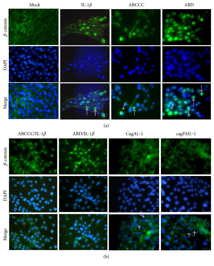
Figure 1.
H. pylori CagA and IL-1β induce β-catenin nuclear translocation. (a) MCF-10A cells were infected with CagA positive strains (ABCCC or ABD) or stimulated with IL-1β. (b) MCF-10A cells were infected with CagA positive strains and stimulated with IL-1β or single infected with CagA negative variants CagA(−) and cagPAI(−). Immunofluorescence images show β-catenin (green) and nuclei (DAPI, blue). Arrows indicate nuclear staining (a) and membrane staining (b) of β-catenin. Figures are representative of three independent experiments performed in duplicate or triplicate.

