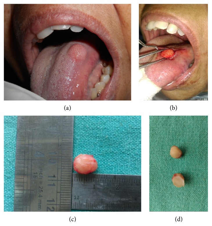
Figure 1.

(a) Intraoral photograph showing well-circumscribed round 1.0 cm × 1.0 cm nodule over dorsum of tongue near left lateral border. (b) Peroperative photograph showing well-circumscribed tumor separated from the surroundings through blunt dissection. (c) Gross specimen appearing well-circumscribed, yellowish white, and oval and measuring 10.0 mm × 5.0 mm × 5.0 mm. (d) Cut surface of the excised tumor appearing homogenous, white, and firm without haemorrhage or necrosis.
