
Figure 2.
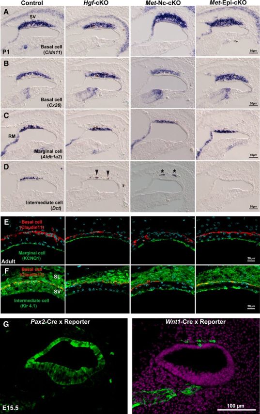
Intermediate cells are absent in the cochlea of Hgf and c-Met mutants at postnatal day 1. A, B, At postnatal day 1, basal cell markers, Claudin11 (Cldn11) and Connexin26 (Cx26), are expressed in the stria vascularis (sv) of Hgf; Pax2-Cre conditional knock-out mouse (Hgf-cKO), neural crest cell-specific c-Met; Wnt1-Cre conditional knock-out mouse (Met-Nc-cKO), and cochlear epithelium-specific c-Met; Pax2-Cre conditional knock-out mouse (Met-Epi-cKO). C, A marginal cell marker, Aldh1a2, is expressed in the absence of HGF–c-MET signaling. D, An intermediate cell/melanocyte marker, Dct, is not observed in the stria vascularis of any of the three conditional mutant mouse lines. Note that a few pigmented cells in the Hgf-cKO may be incorporated into the epithelium (arrowheads) and a few cells remain in the mesenchyme near the cochlear epithelium (Met-Nc-cKO, asterisks). E, Immunofluorescence images show a marginal cell marker, KCNQ1 (green), and a basal cell marker, Claudin11 (red), are expressed in adult stria vascularis. F, An intermediate cell/melanocyte marker, Kir4.1 (green) is not expressed in the stria vascularis (SV) of any of the three conditional mutant mouse lines, while it is expressed in the spiral ligament (SL) of all four genotypes. G, Cre-loxP reporter signals (green) of E15.5 cochlear duct in Pax2-Cre and Wnt1-Cre animals. Note that Pax2-Cre does not exhibit 100% efficiency in the cochlear epithelium. RM, Reissner's membrane.