Abstract
Long-term depression (LTD) and long-term potentiation (LTP) of granule-Purkinje cell synapses are persistent synaptic alterations induced by high and low rises of the intracellular calcium ion concentration ([Ca2+]), respectively. The occurrence of LTD involves the activation of a positive feedback loop formed by protein kinase C, phospholipase A2, and the extracellular signal-regulated protein kinase pathway, and its expression comprises the reduction of the population of synaptic AMPA receptors. Recently, a stochastic computational model of these signalling processes demonstrated that, in single synapses, LTD is probabilistic and bistable. Here, we expanded this model to simulate LTP, which requires protein phosphatases and the increase in the population of synaptic AMPA receptors. Our results indicated that, in single synapses, while LTD is bistable, LTP is gradual. Ca2+ induced both processes stochastically. The magnitudes of the Ca2+ signals and the states of the signalling network regulated the likelihood of LTP and LTD and defined dynamic macroscopic Ca2+ thresholds for the synaptic modifications in populations of synapses according to an inverse Bienenstock, Cooper and Munro (BCM) rule or a sigmoidal function. In conclusion, our model presents a unifying mechanism that explains the macroscopic properties of LTP and LTD from their dynamics in single synapses.
Long-term depression (LTD) and long-term potentiation (LTP) are persistent activity-dependent modifications of the synaptic strength1. One of the best characterized forms of LTD occurs in the synapses between granule cells and Purkinje neurons in the cerebellum1. The induction of LTD involves simultaneous stimulations of parallel fibres and climbing fibres at low frequency (~1 Hz), which promote large elevations of the intracellular calcium ion concentration ([Ca2+])1,2, and activate a positive feedback loop formed by protein kinase C (PKC), cytosolic phospholipase A2 (PLA2), and the extracellular signal-regulated protein kinase (ERK) pathway3,4. LTD expression results from the phosphorylation of synaptic AMPA receptors (AMPARsyn) by PKC5,6, which disrupts their interactions with the glutamate-receptor interacting protein (GRIP)7, and causes their endocytosis8.
Granule-Purkinje cells synapses also exhibit postsynaptic LTP induced by repetitive activations of the parallel fibres at low frequency (1 Hz), which cause low Ca2+ transients1,2. LTP involves the activity of protein phosphatase 1 (PP1), protein phosphatase 2A (PP2A), calcineurin (CaN)9,10, and an increase in the number of AMPARsyn11.
Experimental findings indicated the existence of specific Ca2+ thresholds for the induction of cerebellar synaptic plasticity consistent with the inverse Bienenstock, Cooper and Munro (BCM) rule, which proposes that the postsynaptic strength is potentiated below a sliding modification threshold and depotentiated above it1,12. However, experiments of photolysis of Ca2+-caged compounds demonstrated a sigmoidal correlation between the magnitudes of LTD and the amplitudes of the Ca2+ elevations, but failed to obtain LTP13. A stochastic computational model of this Ca2+-induced LTD demonstrated that its occurrence is probabilistic and modulated by the intensity of the Ca2+ transients used as input signals14. However, a limitation of this earlier model is that it did not simulate mechanisms implicated with LTP, which could reveal a more complex scenario involving Ca2+ and the long-lasting forms of synaptic plasticity.
In this work, we have expanded extensively the previous stochastic computational model of Ca2+-induced LTD to simulate LTP and other molecules implicated with LTD. The additional components of the model included Calmodulin (CaM), Ca2+/Calmodulin protein kinase II α (αCaMKII)15, CaN, Raf kinase inhibitor protein (RKIP), and an endocytic protein (EP) that mediated the internalization of AMPA receptors (AMPARs). Supplementary Table S1 included all the reactions and parameters used in the model and specified which one of them were taken from the previous version. Large-scale computational models of signaling networks are powerful tools for studying the dynamics of the biological systems, but most large-scale models of synaptic plasticity simulate only one process3,16,17. However, the signalling pathways involved with LTP and LTD coexist in the same synapses1,18 and compete for Ca2+ 19. Thus, the aim of our work was to gain insights on the dynamics of the signalling networks involved with the two opposite long-term forms of synaptic plasticity in cerebellum.
Our results showed that, in single synapses, LTD is an all-or-none process, but LTP is graded. Ca2+ transients promoted both processes in a stochastic manner. Nevertheless, the intensity of Ca2+ signals used to induce synaptic plasticity modulated the likelihood of LTP and LTD occurrences in single synapses. In addition, alterations in the components of the signaling network regulated the effects of Ca2+ signals on the induction of LTP and LTD. In consequence, our results indicated the existence of dynamic Ca2+ thresholds for the occurrence of macroscopic synaptic modifications according to the inverse BMC rule proposed for cerebellar plasticity2. Moreover, by limiting the range of magnitudes of Ca2+ transients used as input signals, we obtained the macroscopic sigmoidal relationship observed between the amplitudes of Ca2+ signals and the corresponding levels of depression13 from the inverse BCM rule. With our novel model, we presented a unifying mechanism of opposite forms of postsynaptic long-term synaptic plasticity in Purkinje cells that described their macroscopic characteristics emerging from their elaborated single synapse dynamics.
Results
Stochastic computational model of cerebellar LTD and LTP
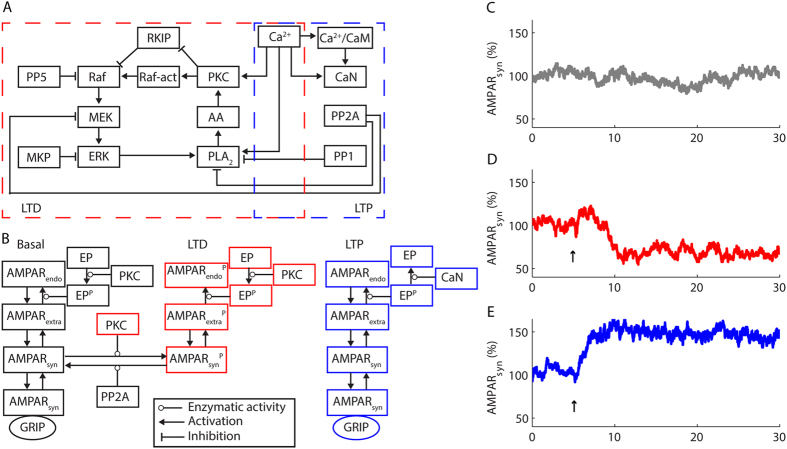
The computational model presented in this work simulated LTD and LTP in a single Purkinje cell dendritic spine, which encloses the signalling machinery of the glutamatergic synapse18,20. We simulated LTD with the positive feedback loop formed by PKC, PLA2, and ERK pathway, which we expanded and updated from a previous version14 based on early models of synaptic plasticity3,16. In our model, Ca2+ elevations transiently activate PKC and PLA214. PKC activates Rapidly accelerated fibrossarcoma (Raf) that activates mitogen-activated ERK kinase (MEK)21. MEK activates ERK21, which phosphorylates PLA2 and sustains its activity after the return of [Ca2+] to its basal level22. PLA2 produces arachidonic acid (AA), a PKC co-factor that activates it synergistically with Ca2+ or alone in high concentrations23. PKC phosphorylates RKIP and contributes to the activation of Raf24. Additionally, PKC regulated ERK pathway through a Raf activator (Raf-act) as implemented previously (Fig. 1A)14. The activation of PKC during LTD caused the synaptic depression through the endocytosis of AMPARs14. In our simulations, AMPARs were constantly trafficked (diffused, endocytosed and reinserted)25 (Fig. 1B,C). However, some synaptic receptors were immobile due to interactions with the scaffold GRIP7. The phosphorylation of AMPARsyn by PKC disrupted these interactions5,7 and promoted their internalization8 (Fig. 1B) and the expression of LTD26,27 (Fig. 1D), which we defined in the model as sustained reductions of the percentage of AMPARsyn. We set the basal percentage of AMPARsyn as 100%.
Figure 1. Stochastic computational model of cerebellar LTP and LTD.
(A,B) Block diagram of the model showing the molecules involved with LTD and LTP (A), and the mechanisms of AMPARs trafficking (B). At rest, we simulated a constant AMPARs trafficking consisted of lateral diffusion from the synapses (AMPARsyn) to extra-synaptic membranes (AMPARextra), from extra-synaptic membranes to endosomes (AMPARendo), and vice-versa. Phosphorylated EP (EPP) catalyzed the internalization of AMPARextra. Part of AMPARsyn interacted with GRIP and did not participate in the constant AMPARs trafficking. During LTD, PKC phosphorylated AMPARsyn (AMPARsynP) and disrupted their interaction with GRIP promoting their internalization. PP2A counteracted PKC action. During LTP, CaN dephosphorylated EPP and blocked AMPARs internalization. PKC counteracted the action of CaN on EP. (C) Simulation of the percentage of AMPARsyn at rest. (D) LTD expression consisted of a persistent reduction of AMPARsyn. (E) During LTP, the model simulated an increase in the percentage of AMPARsyn. The arrows indicate LTD and LTP induction with a Ca2+ pulse of 3 μmol.L−1 and 0.35 μmol.L−1, respectively, and 30 s of duration.
In Purkinje cells, LTP requires protein phosphatases9,10. PP1 and PP2A9, in addition to protein phosphatase 5 (PP5) and the mitogen-activated protein phosphatase (MKP), were included in the signalling network that simulated LTD to counteract the activity of the kinases14. During LTP, these phosphatases prevented the activation of the positive feedback loop PKC-PLA2-ERK pathway. However, to simulate LTP we had to include CaN in the model (Fig. 1A)9,10, which we implemented as a heterodimer composed by a CNA subunit that interacts with Ca2+/CaM, and a CNB subunit with four Ca2+-binding sites28,29.
CaN is implicated in the trafficking of AMPARs30 and plays a pivotal role in endocytosis31. The balance between PKC and CaN controls the phosphorylation of dynamin and syndapin, two proteins involved in vesicles endocytosis31,32. In Purkinje cells, phosphorylated syndapin participates in the endocytosis of AMPARs8. The dephosphorylation of syndapin blocks the internalization of AMPARs8. Thus, we simulated a protein, which we termed EP, that mediated the internalization of AMPARs, but only in its phosphorylated state8. PKC catalysed the phosphorylation of EP8. At rest, the model simulated the constant cycle of AMPARs in and out of synapses25, which requires the basal activity of PKC33 to maintain EP phosphorylated. During LTP, CaN dephosphorylated EP and blocked the endocytosis of AMPARs, without affecting their exocytosis (Fig. 1B). This mechanism of continuous insertion without the concomitant internalization of receptors caused the increase of the AMPARsyn population and the expression of LTP (Fig. 1E). Therefore, the occurrence of LTP in the model involved the persistent increase of the percentage of AMPARsyn.
The role of αCaMKII and phosphatases during the macroscopic occurrence of LTD and LTP
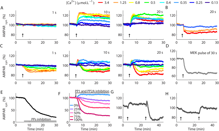
After the implementation of the model, we used Ca2+ pulses with different amplitudes and durations to simulate the photolysis of Ca2+-caged compounds, which can induce LTD13 and LTP2. To compare the results of the simulations with experimental macroscopic curves of LTP and LTD reported in the literature, we used average results of several simulations to represent the responses of populations of synapses. We expected to induce LTP and LTD with Ca2+ pulses of low (~0.3 μmol.L−1) and high amplitudes (>0.5 μmol.L−1), respectively2,13. Pulses of 1 s of duration caused no change in the synaptic strength (Fig. 2A, Supplementary Figs S1 and S2). Longer pulses (10 s and 20 s) promoted LTP for most amplitudes of Ca2+ pulses tested, including high amplitude signals that typically induce LTD in Purkinje cells1,2,13 (Fig. 2A, Supplementary Fig. S1). The blockage of CaN restored the LTD occurrence (Fig. 2B), which indicated that LTP occluded the macroscopic LTD.
Figure 2. Macroscopic curves of LTP and LTD.
(A) In the absence of αCaMKII, high amplitude Ca2+ pulses of 10 s and 20 s resulted in potentiation. (B) The blockage of CaN restored the occurrence of LTD. (C) LTP and LTD in the model with αCaMKII. (D) A pulse of MEK induced LTD. (E) Inhibition of PP1, PP2A and PP5 promoted a slow LTD. (F) Gradual blockages of PP1 and PP2A resulted in LTD induction with a protocol of LTP (Ca2+ pulse of ~0.4 μmol.L−1 and 20 s). (G,H) Reversible synaptic plasticity. In (G), a Ca2+ pulse of ~0.35 μmol.L−1 and 20 s induced LTP. After 20 min, a second pulse of 4 μmol.L−1 and 20 s induced LTD. In (H) we induced weak LTD with a Ca2+ pulse of ~0.9 μmol.L−1 and 1 s, and restored the basal level of AMPARsyn with a Ca2+ pulse of ~0.35 μmol.L−1 and 30 s. Each curve in (A–D) is the average result of 100 runs of the model, and in (E–H) the average of 50 runs. The arrows indicated the occurrence of the Ca2+ pulses. We omitted the standard errors of the mean (SEM) for better visualization. The curves with the mean ± SEM are showed in Supplementary Fig. S2.
Cerebellar LTD requires the activation of the feedback loop PKC-PLA2-ERK3,4, but other molecules are also essential for its occurrence. In αCaMKII knockout mice, protocols of LTD induce LTP in the synapses between granule cells and Purkinje neurons indicating that αCaMKII plays a key role for cerebellar LTD15. Thus, to restore LTD without the blockage of CaN, an evident expansion of the model was the inclusion of αCaMKII, a molecule omitted from most models of cerebellar LTD3,13,14.
αCaMKII has several putative targets during synaptic plasticity, including Raf34,35. Raf activation is a bottleneck for PKC and ERK coupling. Therefore, we implemented αCaMKII acting as a Raf kinase34,35 (Supplementary Fig. S3). The simulation of αCaMKII comprised its detailed binding to Ca2+/CaM and its subsequent autophosphorylation. The autophosphorylation of αCaMKII modulated its affinity for Ca2+/CaM and produced an autonomous state that sustained its partial activity36 in absence of Ca2+/CaM for seconds37, but not for hours as classically thought.
αCaMKII inclusion restored the occurrence of macroscopic LTD induced with high Ca2+ elevations without affecting LTP induction for low Ca2+ transients with prolonged durations (10 s and 20 s) (Fig. 2C). The range of Ca2+ rises necessary to induce LTP in the model (0.15–0.4 μmol.L−1) was similar to experimental estimations (0.1–0.3 μmol.L−1)2. We did not observe LTP for stimulations with Ca2+ pulses of 1 s, which was consequent to the mechanisms of activation of CaN simulated38.
CaN is a heterodimer activated by Ca2+ and Ca2+/CaM28. Under basal [Ca2+], two high affinity Ca2+-binding sites of the regulatory subunit CNB are constantly filled29, but CNA, the subunit that contains the catalytic site of CaN, is inactive28,39,40. The occupancy of the two low affinity Ca2+-binding sites of CNB29,41 during elevations of Ca2+ promotes a conformational change that enables the binding of Ca2+/CaM to CNA and the exposure of its catalytic site39,40,41. Isolated CNA has low catalytic activity, which is stimulated by Ca2+/CaM in absence of CNB42,43. Nevertheless, in the cells CaN always occurs as the heterodimer CNB/CNA41, consequently, its catalytic activation includes the binding of Ca2+ to CNB prior to the binding of Ca2+/CaM to CNA28,29,39. The binding of Ca2+ to CNB occurred with slow rate constants29 in our model and limited the activation of CaN for brief signals38 impairing LTP induction for Ca2+ pulses of 1 s. Consequently, the kinetic aspects of CaN activation constrained the durations of the Ca2+ transients38 that promoted macroscopic LTP (Fig. 2C).
The direction of the synaptic modifications relies on the balance between the activities of protein kinases and phosphatases44. Historically, models of cerebellar LTD implicated the strong inhibition of PP2A by the phosphorylated G-substrate as a key step for synaptic depression3,13. G-substrate is abundant in Purkinje cells and is a putative target for the nitric oxide (NO)-cyclic guanine monophosphate-dependent protein kinase pathway45. However, G-substrate knockout adult mice have normal LTD45. Accordingly, we opted to model LTD without the strong inhibition of PP2A simulated previously3,13. In our model, LTD occurrence involved a shift from a state of low kinase activities at basal [Ca2+], to a state of high kinase activities consequent to the activation of the positive feedback loop. Thus, processes that favour the activation of the loop caused LTD in the model. For instance, a pulse of active MEK promoted LTD4 (Fig. 2D, Supplementary Fig. S4). In addition, the inhibition of PP1, PP5 and PP2A induced a slow LTD46 (Fig. 2E) because it released the inhibition for the activation of the feedback loop PKC-PLA2-ERK. Moreover, partial blockages of PP1 and PP2A promoted the induction of LTD for a LTP protocol (Fig. 2F) as observed experimentally9, and the magnitudes of the depression varied as a function of the levels of phosphatases inhibition (Fig. 2F), which indicated a concentration-dependent effect. Therefore, the existence of mechanisms of LTP and LTD in the same synapses allowed the model to exhibit different outcomes to equivalent protocols as consequences of alterations in the dynamics of its signalling network.
Another implication of the coexistence of signalling mechanisms of LTP and LTD in the same synapses is the possibility of reversibility of plasticity1,2. To verify whether our model could exhibit this property, after the induction of LTP with a low and prolonged Ca2+ transient, we simulated a strong Ca2+ signal and promoted LTD (Fig. 2G). The model also simulated the restoration of the basal synaptic strength after the occurrence of weak LTD (Fig. 2H). However, the model failed to simulate LTP after the induction of strong LTD. The reason for this limitation was the absence of mechanisms to deactivate the positive feedback loop in the model because such mechanisms have not been described. Positive feedback loops promote sustained responses14. Consequently, mechanisms that turn off the positive feedback loop PKC-PLA2-ERK are crucial for the successive occurrences of opposite forms of synaptic plasticity observed experimentally2.
Stochastic induction of graded LTP and bistable LTD in single synapses
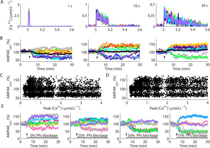
The signalling machinery involved with plasticity in glutamatergic synapses is located in dendritic spines, small structures that act as isolated biochemical compartments20. Each spine encloses a signalling population consisting of few copies of several different molecules18 susceptible to undergo high amplitude stochastic fluctuations in their activities14. Usually, experimental curves of LTP and LTD represent the macroscopic integration of hundreds to thousands of synapses. In the model, we reproduced the macroscopic curves using average results of several simulations of plasticity in single synapses. However, in signalling systems susceptible to stochasticity, the average behaviour can diverge from unitary events14. Thus, the next stage of our work investigated the characteristics of LTP and LTD in single synapses induced by Ca2+ pulses with different amplitudes and durations (Fig. 3A).
Figure 3. Stochastic induction of LTP and LTD in single synapses.
(A,B) Ca2+ pulses of different peak amplitudes and durations (A) induced bistable LTD and graded LTP in the model (B). The black lines are the average results calculated for the 10 runs of the model showed with multiple colours in each panel. The result of each single run for each Ca2+ pulse duration tested is showed with consistent colours in panels A,B. (C) Changes of the percentage of AMPARsyn (measured 25 min after the induction of plasticity) as functions of the peak amplitudes of the Ca2+ pulses (Peak [Ca2+]) used to promote plasticity. (D) Changes of the percentage of AMPARsyn as functions of Peak [Ca2+] used to induce synaptic modification in the model without αCaMKII. Each dot in (C,D) is the result of a single simulation. The durations of the Ca2+ pulses ranged from 1 s to 30 s. (E) Effects of the partial blockages of PP1 and PP2A (termed PPs in the panels) in simulations of single synapses stimulated with a protocol used to induce macroscopic LTP (a single Ca2+ pulse of ~0.35 μmol.L−1 and 20 s of duration).
In single synapses, LTD was an all-or-none process (Fig. 3B), as demonstrated previously14. This bistability resulted from the activation of the positive feedback loop PKC-PLA2-ERK, which promotes robust and persistent responses14. In contrast, LTP happened without mechanisms of self-regulation or amplification. The levels of potentiation resulted from the competition between the activity of CaN and PKC on EP, their common substrate. Consequently, LTP in single synapses was graded (Fig. 3B).
The magnitudes of the Ca2+ pulses did not ensure the occurrence of a specific type of plasticity in single synapses. Ca2+ signals with equivalent peak amplitudes and durations promoted either the occurrence of LTP and LTD, or failed to induce synaptic modifications (Fig. 3A,B, Supplementary Fig. S5). Therefore, the dynamics of plasticity in single synapses diverged from average responses (Fig. 3B, black lines).
The occurrence of opposite forms of plasticity induced by equivalent Ca2+ transients indicated that both LTP and LTD were stochastic processes in the model, which we corroborated assessing the changes of AMPARsyn as a function of the peak Ca2+ rises (Fig. 3C). We measured the alterations of AMPARsyn 25 minutes after the induction of plasticity with Ca2+ pulses of different durations and peak amplitudes. Our results demonstrated a high rate of LTD, measured as reductions of AMPARsyn from its basal value (set as 100%), for the entire range of Ca2+ amplitudes tested. However, LTD predominated as the synaptic modification obtained in the model for stimulations with high amplitude Ca2+ transients (>0.8 μmol.L−1) (Fig. 3C). In contrast, LTP, verified as increases of AMPARsyn from its basal value, happened preferentially for low Ca2+ signals (Fig. 3C).
Next, we addressed whether other components of the model regulated the role of Ca2+ on LTP and LTD inductions. Thus, we verified the changes of AMPARsyn as functions of the peak amplitudes of Ca2+ transients for simulations performed without αCaMKII. The results showed that the absence of αCaMKII increased the range of Ca2+ amplitudes that induced LTP and decreased the occurrence of LTD (Fig. 3D). This increase of LTP occurrence combined with the reduction of LTD occurrence promoted the macroscopic curves of plasticity that failed to exhibit macroscopic depression showed previously (Fig. 2A).
Simulations of plasticity in single synapses induced with a LTP protocol in the presence of partial blockages of PP1 and PP2A indicated that the reduction of phosphatases activities increased the occurrence of microscopic LTD in a concentration-dependent manner (Fig. 3E), and promoted the macroscopic curves with different magnitudes of depression showed in Fig. 2F. Thus, while the absence of αCaMKII increased the induction of LTP (Fig. 3D) in comparison to the control model (Fig. 3C) and disrupted the occurrence of macroscopic LTD (Fig. 2A), reduction of the activities of PP1 and PP2A increased LTD occurrence in single synapses (Fig. 3E) and promoted macroscopic curves of depression for protocols that should induce LTP (Fig. 2F). Historically, the discrimination between the induction of LTP and LTD is attributed to the existence of specific Ca2+ thresholds, which would activate Ca2+-dependent kinases and phosphatases with distinct Ca2+-affinities19. Our results demonstrated that different magnitudes of Ca2+ signals modulate the stochastic induction of LTP and LTD, but this modulation was not fixed. Changes in the components of the model regulated the role of Ca2+ on the induction of LTP and LTD in a dynamic manner.
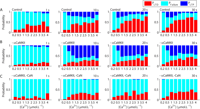
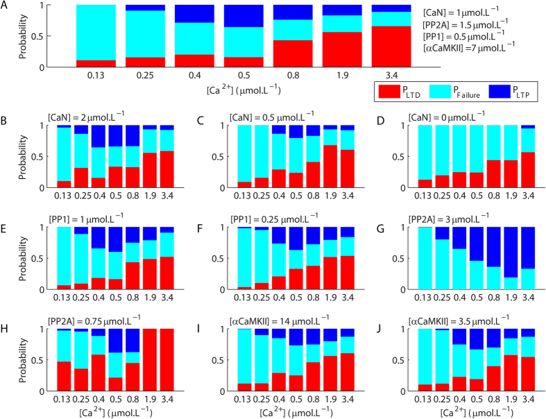
To quantify the role of Ca2+ signals on the induction of LTP and LTD, we calculated the probability of unitary occurrences of LTD (PLTD), LTP (PLTP) and failure of plasticity (PFailure) for Ca2+ pulses with different amplitudes and durations. Our results showed that PLTD increased with the increment of the durations and peak amplitudes of the Ca2+ signals used to trigger plasticity (Fig. 4A)14. PLTP was low for short Ca2+ pulses (1 s). For Ca2+ signals of 10 s, PLTP was low for weak Ca2+ rises, increased for amplitudes ranging from 1–3.5 μmol.L−1 and dropped for higher concentrations while PLTD increased progressively with the increment of the amplitudes of the Ca2+ signals (Fig. 4A). For pulses of 20–30 s of duration, PLTP was high for Ca2+ signals with low peak amplitudes (0.5–1 μmol.L−1), and decreased progressively with the increment of the amplitudes of the pulses, which caused the increase of PLTD (Fig. 4A). Taken together, these results suggested that the activation of the Ca2+-dependent molecules involved with both LTP and LTD increased with the intensification of the magnitudes of the Ca2+ signals. However, because cerebellar LTD involves the activation of a positive feedback loop, which produces sustained patterns of activation14, its occurrence occluded LTP. To investigate this hypothesis, we calculated PLTD, PLTP and PFailure for modified versions of the model. Simulations performed in absence of αCaMKII presented a clear reduction of PLTD in comparison with the control model and higher PLTP for all magnitudes of Ca2+ signals tested (Fig. 4B). In contrast, simulations of the model in the absence of αCaMKII and CaN (Fig. 4C) had no LTP, but exhibited PLTD similar to the values observed for the model without αCaMKII (Fig. 4B). These results indicated that the occurrence of LTP did not interfere with PLTD in the conditions tested, but LTD occurrences altered PLTP. Therefore, the components of the model affected in a non-linear manner the occurrences of unitary LTP and LTD and dynamically regulated the role of Ca2+ on the stochastic induction of the opposite forms of synaptic plasticity.
Figure 4. Probabilistic induction of long-term synaptic plasticity.
(A) The durations and peak amplitudes of the Ca2+ pulses regulated the probabilities of induction of LTP (PLTP), LTD (PLTD), and failure of induction of synaptic plasticity (PFailure) in the control model. The regulatory role of Ca2+ on PLTP and PLTD was further modulated by the absence of αCaMKII (B), which promoted a global reduction of PLTD and an increase of PLTP. The simultaneous absence of αCaMKII and CaN had no additional effect on PLTD, but suppressed the occurrence of LTP (C). For each Ca2+ concentration in each panel, we analysed the results of 50–200 single runs of the model.
Macroscopic Ca2+ thresholds to induce LTD and LTP
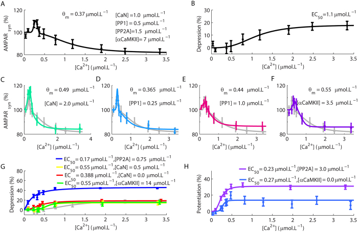
The modulatory role of Ca2+ transients in the directions of synaptic plasticity in single synapses determined macroscopic Ca2+ thresholds for the induction of LTD and LTP (Fig. 5A, Supplementary Fig. S6). These results were observed with Ca2+ pulses of 10–30 s. In Fig. 5A, the first Ca2+ threshold, observed for low amplitude Ca2+ transients, represented the change from non-plasticity to LTP, and the second was the threshold for the conversion of LTP to LTD, which corresponds to the crossover point (θm) predicted in the classical BCM rule12. Thus, the macroscopic curves of plasticity obtained with our model corroborated the existence of Ca2+ thresholds for synaptic modifications consistent with the inverse BMC rule2.
Figure 5. Macroscopic Ca2+ thresholds for the occurrences of synaptic modifications.
(A,B) An inverse BCM rule (A) and a sigmoid function (B) described the correlation between the macroscopic synaptic modifications and the magnitudes of Ca2+ pulses according to the range of Ca2+ amplitudes analysed. The term EC50 stands for the [Ca2+] required to achieve half-maximum LTD. (C) Simulations performed with higher [CaN] (the control concentration of CaN and other components of the model are indicated in (A)). (D,E) A reduction of [PP1] (D) had little effect on the overall behaviour of the model, but an increase (E) in its concentration changed θm. (F) Reduction of [αCaMKII] altered the value of θm of the inverse BMC rule. In (C–F) the light gray line corresponds to the results obtained with the control model (A) replotted for comparison. (G) An increase of [αCaMKII] or a reduction of [CaN] or [PP2A] suppressed the occurrence of LTP (the control result (B) was replotted in gray for comparison). (H) The elevation of [PP2A] or the absence of αCaMKII resulted in the suppression of LTD. Each dot in the panels is the mean + SEM calculated for 100 simulations. The duration of the Ca2+ pulses was 20 s for all simulations.
Interestingly, a previous work reported the existence of a macroscopic sigmoidal relationship between the magnitudes of LTD and the levels of Ca2+ rises, but failed to observe LTP13. However, the range of [Ca2+] investigated in this work varied from approximately 0.5 to 6 μmol.L−1 13. In our work, we observed macroscopic LTP only for Ca2+ transients lower than 0.5 μmol.L−1 (θm = 0.37 μmol.L−1), which is consistent with other experimental results2. Nevertheless, by removing the results obtained for Ca2+ pulses below from 0.5 μmol.L−1 from our analyses, we obtained the same sigmoid function observed experimentally13 (Fig. 5B). Therefore, our results supported the existence of the two macroscopic rules (the inverse BCM rule and the sigmoid relationship) to describe the relations between the levels of Ca2+ rise and the occurrence of opposite forms of synaptic plasticity.
In the simulations of single synapses described previously (Figs 3 and 4), we verified that different components of the model affected the occurrences of plasticity. These results suggested that the components of the model regulate the macroscopic Ca2+ thresholds for LTP and LTD. To investigate this aspect of the model, we varied the concentrations of some of its components and verified their impacts on the macroscopic Ca2+ thresholds for the induction of LTP and LTD (Fig. 5C–H, Supplementary Fig. S7). Simulations with a higher concentration of CaN ([CaN] = 2 μmol.L−1, the control concentration was 1 μmol.L−1) resulted in stronger LTP and affected θm (Fig. 5C). A reduction of PP1 concentration ([PP1] = 0.25 μmol.L−1, in the control model [PP1] = 0.5 μmol.L−1) had no effect on the Ca2+ thresholds for LTP and LTD, but an increase of [PP1] (1 μmol.L−1) altered θm (Fig. 5D,E). We also observed an alteration of θm for simulations with a reduced αCaMKII concentration ([αCaMKII] = 3.5 μmol.L−1, [αCaMKII] was ~7 μmol.L−1 in the control model) (Fig. 5F). In contrast, an increase of [αCaMKII] (14 μmol.L−1) suppressed LTP and promoted a sigmoidal relation between the amplitudes of the Ca2+ signals and the magnitudes of LTD (Fig. 5G). We verified similar results for simulations with reduced [CaN] (0.5 and 0 μmol.L−1) and reduced concentration of PP2A ([PP2A] = 0.75 μmol.L−1, its control concentration was 1.5 μmol.L−1) (Fig. 5G). Additionally, all these curves (Fig. 5G) exhibited lower Ca2+ requirement to achieve half-maximum depression (EC50) in comparison to the control model (Fig. 5G light gray line). We suppressed LTD and obtained sigmoidal relations between the amplitudes of the Ca2+ signals and the magnitudes of LTP by setting [αCaMKII] as 0 μmol.L−1 or increasing [PP2A] (from 1.5 μmol.L−1 to 3 μmol.L−1) (Fig. 5H). Therefore, alterations of the molecules involved with synaptic plasticity affected the thresholds and the rules that associate changes of [Ca2+] with the directions of the macroscopic forms of synaptic plasticity.
Microscopically, the curves presented in Fig. 5 emerged from the combinations of the probabilities of LTP and LTD occurrences and the probability of failure of synaptic plasticity. The BCM rule observed for the control model (Fig. 5A) resulted from the balance of PLTP, PLTD and PFailure that changed with the amplitudes of the Ca2+ signals used to induce plasticity (Fig. 6A). Modifications of the components of the model that promoted alterations in the rules and thresholds for the macroscopic forms of synaptic plasticity did so by affecting the balances between PLTP, PLTD and PFailure. The increase of [CaN] that promoted stronger LTP and affected θm in Fig. 5C resulted from an overall increase of PLTP and from alterations in the peak amplitudes of the Ca2+ signals associated with maximum PLTP (Fig. 6B). Decreases of [CaN] had the opposite effect (Fig. 6C,D). The increase of [PP1] caused an enhancement of PLTP for some amplitudes of Ca2+ transients (Fig. 6E), and the decrease of [PP1] had little effect on both PLTP and PLTD (Fig. 6F), which is consistent with the fact that this alteration had only slight effects on the Ca2+ thresholds for macroscopic LTP and LTD occurrences (Fig. 5D). In contrast, the increase of [PP2A] suppressed the occurrence of unitary LTD (PLTD = 0). As a result, the balance between PFailure and PLTP (Fig. 6G) promoted a sigmoid function between the levels of Ca2+ rises and the magnitudes of macroscopic LTP (Fig. 5H). The reduction of [PP2A] had the opposite effect and decreased PLTP and enhanced PLTD (Fig. 6H). The increase of [αCaMKII], which blocked the occurrences of macroscopic LTP (Fig. 5G), did not supress the occurrence of unitary LTP, but decreased PLTP (Fig. 6I). However, the decrease of [αCaMKII] altered PLTP and PLTD for intermediary peak amplitudes of the Ca2+ signals (Fig. 6J) in comparison to the control model and, in consequence, affected θm of the macroscopic LTP and LTD occurrences (Fig. 5F). Thus, the macroscopic relations between Ca2+ and the inductions of LTP and LTD are dynamically regulated by changes in the probabilities of unitary occurrences of synaptic plasticity.
Figure 6. Modulations of the probabilities of LTP and LTD inductions.
(A) Probabilities of LTP (PLTP), LTD (PLTD), and failure (PFailure) of inductions of synaptic plasticity for the control model stimulated with Ca2+ pulses of 20 s of duration and different peak concentrations. (B–J) PLTP, PLTP, and PFailure obtained for Ca2+ pulses of 20 s for modified versions of the model with an increase of [CaN] (B), decrease of [CaN] (C), absence of CaN (D), increase of [PP1] (E), decrease of [PP1] (F), increase of [PP2A] (G), decrease of [PP2A] (H), increase of [αCaMKII] (I), and decrease of [αCaMKII] (J). For each Ca2+ concentration in each panel, we analysed the results of 50–100 single runs of the model.
Discussion
We presented a unifying model of postsynaptic cerebellar LTP and LTD in Purkinje cells. There are few other models of the signalling mechanisms of synaptic plasticity in the cerebellum, and most of them focused only on LTD and were solved deterministically3,13,47. The first stochastic model of LTD is recent and indicated that stochasticity plays a central role in the macroscopic curves of plasticity14. In this work, we have expanded and updated the stochastic model of LTD and incorporated mechanisms to simulate LTP. The model reproduced several properties of LTP and LTD observed experimentally, and correlated them with the dynamics of plasticity in single synapses. In this way, the model provided a unified mechanistic explanation for many experimental observations of LTP and LTD occurrences in granule-Purkinje cell synapses. Still, the model has important limitations. For instance, experimental findings reported that both NO and derivatives of AA are involved with LTP and LTD11,48, and CaMKII regulates NO at least during the depression47, but these processes were not implemented in our model. Nevertheless, our work revealed new aspects of the dynamics of LTD and LTP that are testable experimentally.
Historically, the balance between the activity of protein kinases and phosphatases regulated by Ca2+ is considered the key element for the discrimination between the occurrences of LTP and LTD19,44. This observation was initially proposed for the synapses between CA3 and CA1 hippocampal pyramidal neurons19. Hippocampal LTP involves the activation of αCaMKII, and LTD requires CaN19,44. Both CaN and αCaMKII are activated by Ca2+/CaM39,41,49, but CaN has a 1000-fold higher affinity for Ca2+/CaM in comparison to αCaMKII (~12 pmol.L−1 50 for CaN and ~4–20 nmol.L−1 for αCaMKII, which corresponds to its affinity measured in presence of nucleotides51,52,53). Hippocampal LTD and LTP require low and high Ca2+ rises, respectively54. Consequently, a central hypothesis to explain the direction of the synaptic plasticity explored in many computational models17,55 relies on the differences between the affinities of CaN and αCaMKII for Ca2+/CaM, which would promote their differential activations for the low and high Ca2+ elevations required for hippocampal LTD and LTP induction, respectively19. However, our results indicated that the extrapolation of this idea to the discrimination between LTP and LTD for different levels of Ca2+ rises in Purkinje cells is an oversimplification.
In this work, unitary occurrences of LTP and LTD were stochastic processes. LTD was bistable, but LTP was graded. Due to the probabilistic nature of unitary LTP and LTD, the amplitudes of Ca2+ elevations used as input signals did not ensure the occurrence of any particular type of plasticity and played only a modulatory role. Additionally, both LTP and LTD were highly modulated by other signalling species of the model. Thus, not only CaN and αCaMKII, but also several other components of the model affected the probabilities of unitary occurrences of LTP and LTD. Consequently, our results support the existence of dynamic rather than static macroscopic Ca2+-thresholds for the occurrences of LTP and LTD.
Materials and Methods
We built the computational model of LTP and LTD using BioNetGen56, a rule-based software for modelling biochemical networks. We solved the simulations stochastically with the SSA algorithm.
The model consisted of a well-mixed compartment containing mechanisms of Ca2+ dynamics, the signalling network involved with LTP and LTD, and AMPARs trafficking. The detailed descriptions of the components of the model with their respective parameters (Supplementary Table S1), references, validations (Supplementary Fig. S8), and additional analyses (Supplementary Fig. S9) are given in the Supplementary Methods.
Most simulations modelled a time interval of 37 minutes; the first seven minutes comprised the period necessary for the system to reach steady-state and were withdrawn from the analyses. The time course analysed included an initial interval of five minutes before the inductions of plasticity plus 25 minutes, a temporal interval in which the activation of the feedback loop is essential4. In Figs 3C,D and 5, we measured the percentage of AMPARsyn 25 minutes after the induction of plasticity. In Fig. 5, the dots in each curve are means ± standard error of the mean (SEM) calculated for 100 simulations.
We fitted the curves of Fig. 5A,C–F using an equation given as follows:
 |
where a is a scaling factor, b refers to the [Ca2+] required for maximum LTP, c stands for the width of the Gaussian curve for LTP occurrence, LTDmax refers to the maximum depression, nHill is the Hill coefficient and EC50 is the [Ca2+] required to induce half-maximum depression.
The sigmoid function used to fit the curves of Fig. 5B,G is described as (15):
 |
We used the same Equation (2) to fit the sigmoid curves of Fig. 5H replacing LTD for LTP. We fitted all curves in Fig. 5 using the Matlab Curve Fitting Tool (cftool) with 95% of confidence interval.
To calculate the probabilities of LTP and LTD induction in Figs 4 and 6, we measured the percentage of AMPARsyn 25 minutes after the induction of synaptic plasticity for single runs of the model stimulated with Ca2+ pulses with different durations and peak amplitudes. We defined increases of AMPARsyn of 20% and above as LTP and reductions of AMPARsyn of 20% and below as LTD. Variations of the population of AMPARsyn between 80–120% (the standard population was set as 100%) were attributed to the stochasticity of the model and treated as failures of synaptic plasticity induction.
Additional Information
How to cite this article: Antunes, G. et al. Stochastic Induction of Long-Term Potentiation and Long-Term Depression. Sci. Rep. 6, 30899; doi: 10.1038/srep30899 (2016).
Supplementary Material
Acknowledgments
Research supported by the IRTG 1740/TRP 2015/50122-0 funded by DFG/FAPESP, FAPESP grants (2014/08481-0 and 2013/07699-0) and CNPq grant (306251/2014-0).
Footnotes
Author Contributions G.A. designed research, built the model, performed the simulations, analysed the data and wrote the manuscript. F.M.S.-d.-S. designed research, performed the simulations, analysed the data and wrote the manuscript, A.C.R. wrote the manuscript.
References
- Jörntell H. & Hansel C. Synaptic memories upside down: bidirectional plasticity at cerebellar parallel fiber-Purkinje cell synapses. Neuron 52, 227–238 (2006). [DOI] [PubMed] [Google Scholar]
- Coesmans M., Weber J. T., De Zeeuw C. I. & Hansel C. Bidirectional parallel fiber plasticity in the cerebellum under climbing fiber control. Neuron 44, 691–700 (2004). [DOI] [PubMed] [Google Scholar]
- Kuroda S., Schweighofer N. & Kawato M. Exploration of signal transduction pathways in cerebellar long-term depression by kinetic simulation. J Neurosci 21, 5693–5702 (2001). [DOI] [PMC free article] [PubMed] [Google Scholar]
- Tanaka K. & Augustine G. J. A positive feedback signal transduction loop determines timing of cerebellar long-term depression. Neuron 59, 608–620 (2008). [DOI] [PMC free article] [PubMed] [Google Scholar]
- Chung H. J., Steinberg J. P., Huganir R. L. & Linden D. J. Requirement of AMPA receptor GluR2 phosphorylation for cerebellar long-term depression. Science 300, 1751–1755 (2003). [DOI] [PubMed] [Google Scholar]
- Matsuda S., Mikawa S. & Hirai H. Phosphorylation of serine-880 in GluR2 by protein kinase C prevents its C terminus from binding with glutamate receptor-interacting protein. J Neurochem 73, 1765–1768 (1999). [DOI] [PubMed] [Google Scholar]
- Matsuda S., Launey T., Mikawa S. & Hirai H. Disruption of AMPA receptor GluR2 clusters following long-term depression induction in cerebellar Purkinje neurons. EMBO J 19, 2765–2774 (2000). [DOI] [PMC free article] [PubMed] [Google Scholar]
- Anggono V. et al. PICK1 interacts with PACSIN to regulate AMPA receptor internalization and cerebellar long-term depression. Proc Natl Acad Sci USA 110, 13976–13981 (2013). [DOI] [PMC free article] [PubMed] [Google Scholar]
- Belmeguenai A. & Hansel C. A role for protein phosphatases 1, 2A, and 2B in cerebellar long-term potentiation. J Neurosci 25, 10768–10772 (2005). [DOI] [PMC free article] [PubMed] [Google Scholar]
- Schonewille M. et al. Purkinje cell-specific knockout of the protein phosphatase PP2B impairs potentiation and cerebellar motor learning. Neuron 67, 618–628 (2010). [DOI] [PMC free article] [PubMed] [Google Scholar]
- Kakegawa W. & Yuzaki M. A mechanism underlying AMPA receptor trafficking during cerebellar long-term potentiation. Proc Natl Acad Sci USA 102, 17846–17851 (2005). [DOI] [PMC free article] [PubMed] [Google Scholar]
- Bienenstock E. L., Cooper L. N. & Munro P. W. Theory for the development of neuron selectivity: orientation specificity and binocular interaction in visual cortex. J Neurosci 2, 32–48 (1982). [DOI] [PMC free article] [PubMed] [Google Scholar]
- Tanaka K. et al. Ca2+ requirements for cerebellar long-term synaptic depression: role for a postsynaptic leaky integrator. Neuron 54, 787–800 (2007). [DOI] [PubMed] [Google Scholar]
- Antunes G. & De Schutter E. A stochastic signaling network mediates the probabilistic induction of cerebellar long-term depression. J Neurosci 32, 9288–9300 (2012). [DOI] [PMC free article] [PubMed] [Google Scholar]
- Hansel C. et al. alphaCaMKII is essential for cerebellar LTD and motor learning. Neuron 51, 835–843 (2006). [DOI] [PubMed] [Google Scholar]
- Bhalla U. S. & Iyengar R. Emergent properties of networks of biological signaling pathways. Science 283, 381–387 (1999). [DOI] [PubMed] [Google Scholar]
- Kotaleski J. H. & Blackwell K. T. Modelling the molecular mechanisms of synaptic plasticity using systems biology approaches. Nat Rev Neurosci 11, 239–251 (2010). [DOI] [PMC free article] [PubMed] [Google Scholar]
- Cheng D. et al. Relative and absolute quantification of postsynaptic density proteome isolated from rat forebrain and cerebellum. Mol Cell Proteomics 5, 1158–1170 (2006). [DOI] [PubMed] [Google Scholar]
- Lisman J. A mechanism for the Hebb and the anti-Hebb processes underlying learning and memory. Proc Natl Acad Sci USA 86, 9574–9578 (1989). [DOI] [PMC free article] [PubMed] [Google Scholar]
- Harris K. M. & Stevens J. K. Dendritic spines of rat cerebellar Purkinje cells: serial electron microscopy with reference to their biophysical characteristics. J Neurosci 8, 4455–4469 (1988). [DOI] [PMC free article] [PubMed] [Google Scholar]
- Pearson G. et al. Mitogen-activated protein (MAP) kinase pathways: regulation and physiological functions. Endocr Rev 22, 153–183 (2001). [DOI] [PubMed] [Google Scholar]
- Das S., Rafter J. D., Kim K. P., Gygi S. P. & Cho W. Mechanism of group IVA cytosolic phospholipase A(2) activation by phosphorylation. J Biol Chem 278, 41431–41442 (2003). [DOI] [PubMed] [Google Scholar]
- O’Flaherty J. T., Chadwell B. A., Kearns M. W., Sergeant S. & Daniel L. W. Protein kinases C translocation responses to low concentrations of arachidonic acid. J Biol Chem 276, 24743–24750 (2001). [DOI] [PubMed] [Google Scholar]
- Yamamoto Y. et al. Raf kinase inhibitory protein is required for cerebellar long-term synaptic depression by mediating PKC-dependent MAPK activation. J Neurosci 32, 14254–14264 (2012). [DOI] [PMC free article] [PubMed] [Google Scholar]
- Czöndör K. et al. Unified quantitative model of AMPA receptor trafficking at synapses. Proc Natl Acad Sci USA 109, 3522–3527 (2012). [DOI] [PMC free article] [PubMed] [Google Scholar]
- Linden D. J. The expression of cerebellar LTD in culture is not associated with changes in AMPA-receptor kinetics, agonist affinity, or unitary conductance. Proc Natl Acad Sci USA 98, 14066–14071 (2001). [DOI] [PMC free article] [PubMed] [Google Scholar]
- Wang Y. T. & Linden D. J. Expression of cerebellar long-term depression requires postsynaptic clathrin-mediated endocytosis. Neuron 25, 635–647 (2000). [DOI] [PubMed] [Google Scholar]
- Stemmer P. M. & Klee C. B. Dual calcium ion regulation of calcineurin by calmodulin and calcineurin B. Biochemistry 33, 6859–6866 (1994). [DOI] [PubMed] [Google Scholar]
- Feng B. & Stemmer P. M. Interactions of calcineurin A, calcineurin B, and Ca2+. Biochemistry 38, 12481–12489 (1999). [DOI] [PubMed] [Google Scholar]
- Beattie E. C. et al. Regulation of AMPA receptor endocytosis by a signaling mechanism shared with LTD. Nat Neurosci 3, 1291–1300 (2000). [DOI] [PubMed] [Google Scholar]
- Liu J. P., Sim A. T. & Robinson P. J. Calcineurin inhibition of dynamin I GTPase activity coupled to nerve terminal depolarization. Science 265, 970–973 (1994). [DOI] [PubMed] [Google Scholar]
- Anggono V. et al. Syndapin I is the phosphorylation-regulated dynamin I partner in synaptic vesicle endocytosis. Nat Neurosci 9, 752–760 (2006). [DOI] [PMC free article] [PubMed] [Google Scholar]
- Tatsukawa T., Chimura T., Miyakawa H. & Yamaguchi K. Involvement of basal protein kinase C and extracellular signal-regulated kinase 1/2 activities in constitutive internalization of AMPA receptors in cerebellar Purkinje cells. J Neurosci 26, 4820–4825 (2006). [DOI] [PMC free article] [PubMed] [Google Scholar]
- Illario M. et al. Calcium/calmodulin-dependent protein kinase II binds to Raf-1 and modulates integrin-stimulated ERK activation. J Biol Chem 278, 45101–45108 (2003). [DOI] [PubMed] [Google Scholar]
- Salzano M. et al. Calcium/calmodulin-dependent protein kinase II (CaMKII) phosphorylates Raf-1 at serine 338 and mediates Ras-stimulated Raf-1 activation. Cell Cycle 11, 2100–2106 (2012). [DOI] [PubMed] [Google Scholar]
- Coultrap S. J., Buard I., Kulbe J. R., Dell’Acqua M. L. & Bayer K. U. CaMKII autonomy is substrate-dependent and further stimulated by Ca2+/calmodulin. J Biol Chem 285, 17930–17937 (2010). [DOI] [PMC free article] [PubMed] [Google Scholar]
- Lee S. J., Escobedo-Lozoya Y., Szatmari E. M. & Yasuda R. Activation of CaMKII in single dendritic spines during long-term potentiation. Nature 458, 299–304 (2009). [DOI] [PMC free article] [PubMed] [Google Scholar]
- Antunes G., Roque A. C. & Simoes de Souza F. M. Modelling intracellular competition for calcium: kinetic and thermodynamic control of different molecular modes of signal decoding. Scientific Reports 6, 23730 (2016). [DOI] [PMC free article] [PubMed] [Google Scholar]
- Shen X. et al. The secondary structure of calcineurin regulatory region and conformational change induced by calcium/calmodulin binding. J Biol Chem 283, 11407–11413 (2008). [DOI] [PubMed] [Google Scholar]
- Yang S. A. & Klee C. B. Low affinity Ca2+-binding sites of calcineurin B mediate conformational changes in calcineurin A. Biochemistry 39, 16147–16154 (2000). [DOI] [PubMed] [Google Scholar]
- Klee C. B., Ren H. & Wang X. Regulation of the calmodulin-stimulated protein phosphatase, calcineurin. J Biol Chem 273, 13367–13370 (1998). [DOI] [PubMed] [Google Scholar]
- Perrino B. A., Ng L. Y. & Soderling T. R. Calcium regulation of calcineurin phosphatase activity by its B subunit and calmodulin. Role of the autoinhibitory domain. J Biol Chem 270, 7012 (1995). [DOI] [PubMed] [Google Scholar]
- Merat D. L., Hu Z. Y., Carter T. E. & Cheung W. Y. Bovine brain calmodulin-dependent protein phosphatase. Regulation of subunit A activity by calmodulin and subunit B. J. Biol. Chem. 260, 11053–11059 (1985). [PubMed] [Google Scholar]
- Woolfrey K. M. & Dell’Acqua M. L. Coordination of Protein Phosphorylation and Dephosphorylation in Synaptic Plasticity. J Biol Chem, doi: 10.1074/jbc.R115.657262 (2015) [DOI] [PMC free article] [PubMed] [Google Scholar]
- Endo S. et al. Dual involvement of G-substrate in motor learning revealed by gene deletion. Proc Natl Acad Sci USA 106, 3525–3530 (2009). [DOI] [PMC free article] [PubMed] [Google Scholar]
- Eto M., Bock R., Brautigan D. L. & Linden D. J. Cerebellar long-term synaptic depression requires PKC-mediated activation of CPI-17, a myosin/moesin phosphatase inhibitor. Neuron 36, 1145–1158 (2002). [DOI] [PubMed] [Google Scholar]
- Kawaguchi S. Y. & Hirano T. Gating of long-term depression by Ca2+/calmodulin-dependent protein kinase II through enhanced cGMP signalling in cerebellar Purkinje cells. J Physiol 591, 1707–1730 (2013). [DOI] [PMC free article] [PubMed] [Google Scholar]
- Wang D. J. et al. Long-term potentiation at cerebellar parallel fiber-Purkinje cell synapses requires presynaptic and postsynaptic signaling cascades. J Neurosci 34, 2355–2364 (2014). [DOI] [PMC free article] [PubMed] [Google Scholar]
- Stratton M. M., Chao L. H., Schulman H. & Kuriyan J. Structural studies on the regulation of Ca2+/calmodulin dependent protein kinase II. Curr Opin Struct Biol 23, 292–301 (2013). [DOI] [PMC free article] [PubMed] [Google Scholar]
- Quintana A. R., Wang D., Forbes J. E. & Waxham M. N. Kinetics of calmodulin binding to calcineurin. Biochem Biophys Res Commun 334, 674–680 (2005). [DOI] [PubMed] [Google Scholar]
- Forest A. et al. Role of the N- and C-lobes of calmodulin in the activation of Ca(2+)/calmodulin-dependent protein kinase II. Biochemistry 47, 10587–10599 (2008). [DOI] [PMC free article] [PubMed] [Google Scholar]
- Török K., Tzortzopoulos A., Grabarek Z., Best S. L. & Thorogate R. Dual effect of ATP in the activation mechanism of brain Ca(2+)/calmodulin-dependent protein kinase II by Ca(2+)/calmodulin. Biochemistry 40, 14878–14890 (2001). [DOI] [PubMed] [Google Scholar]
- Tzortzopoulos A. & Török K. Mechanism of the T286A-mutant alphaCaMKII interactions with Ca2+/calmodulin and ATP. Biochemistry 43, 6404–6414 (2004). [DOI] [PubMed] [Google Scholar]
- Yang S. N., Tang Y. G. & Zucker R. S. Selective induction of LTP and LTD by postsynaptic [Ca2+]i elevation. J Neurophysiol 81, 781–787 (1999). [DOI] [PubMed] [Google Scholar]
- Stefan M. I., Edelstein S. J. & Le Novère N. An allosteric model of calmodulin explains differential activation of PP2B and CaMKII. Proc Natl Acad Sci USA 105, 10768–10773 (2008). [DOI] [PMC free article] [PubMed] [Google Scholar]
- Faeder J. R., Blinov M. L. & Hlavacek W. S. Rule-based modeling of biochemical systems with BioNetGen. Methods Mol Biol 500, 113–167 (2009). [DOI] [PubMed] [Google Scholar]
Associated Data
This section collects any data citations, data availability statements, or supplementary materials included in this article.


