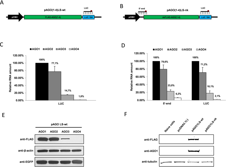
Figure 1. Different overexpression efficiency of human Argonaute coding sequences.
(A) Structure of pAGO1LS-wt, pAGO2LS-wt, pAGO3LS-wt and pAGO4LS-wt used for transient overexpression of recombinant human Argonautes coding regions containing N-terminal FLAG-tag. The location of the luciferase TaqMan qPCR probe (LUC) is indicated on the plasmid map. (B) Structure of pAGO1LS-nk, pAGO2LS-nk, pAGO3LS-nk and pAGO4LS-nk encoding protein synthesis-impaired AGO cDNAs (lacking Kozak sequences and the first 2 nucleotides in the starting ATG codons). The location of the 5′-end and 3′-end (LUC) TaqMan qPCR probes is indicated on the plasmid map. TaqMan qRT-PCR analysis was used to estimate the relative overexpression of recombinant Argonaute transcripts generated from pAGO(1–4)LS-wt (C) and pAGO(1–4)LS-nk plasmids (D) in U2OS cells 24 h after transient transfection. The qPCR data is presented as mRNA levels relative to cells transfected with AGO1 encoding plasmids, and normalized on the NeoR mRNA levels in each sample. Each bar represents mean + S.D of three independent transfections. (E) Western immunoblotting demonstrating expression of FLAG-AGO1, FLAG-AGO2, FLAG-AGO3, FLAG-AGO4, EGFP and β-actin proteins in U2OS cells 24 h after transfection with pAGO1LS-wt, pAGO2LS-wt, pAGO3LS-wt and pAGO4LS-wt vectors mixed with equal amount of pEFGP-C1 plasmid. (F) Western immunoblotting performed with anti-FLAG and anti-AGO1 antibodies demonstrating the absence of recombinant AGO1 protein production in U2OS cells after transfection with pAGO1LS-nk plasmid, in contrast to cells transfected with pAGO1LS-wt.

