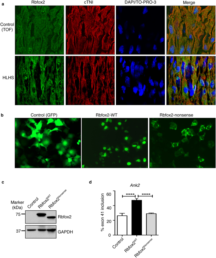
Figure 2. Subcellular distribution of Rbfox2 is altered in HLHS right ventricles.
(a) Immunofluorescence (IF) of Rbfox2 in right ventricle sagittal sections of infants with HLHS or Tetralogy of Fallot (control). Cardiomyocytes were marked with cardiac troponin I. Nuclei were stained with DAPI/TO-PRO-3. Fluorescence images were obtained at 80X magnifications with a confocal laser-scanning microscope (LSM 510META, Carl Zeiss) at the University of Texas Medical Branch imaging core facility. (b) Representative images of GFP (control), GFP-tagged wild type (Rbfox2WT) and nonsense mutant (Rbfox2Nonsense) of Rbfox2 protein in COS M6 cells. (c) WB analysis of GFP-Rbfox2WT and GFP-Rbfox2Nonsense protein in COS M6 cells using anti-Rbfox2 antibody. GAPDH WB was used as a loading control. (d) Percent inclusion of Ank2 exon 41 in control (vector), GFP-Rbfox2WT or Rbfox2Nonsense mutant expressing COS M6 cells determined by qRT-PCR (n ≥ 3). Each reaction was done as triplicates for 3 different experiments and significance was calculated using one-way ANOVA.

