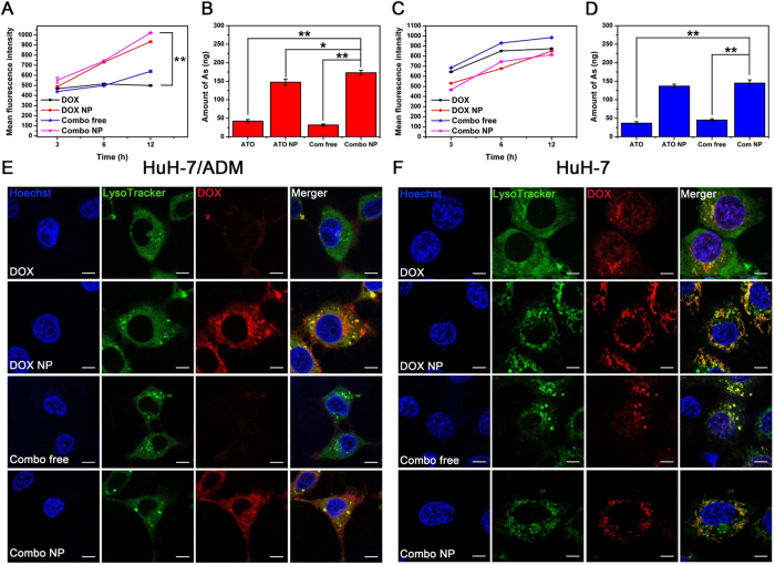
Figure 3. Cellular drug accumulation and localization.
Flow cytometry analysis of DOX fluorescence in (A) HuH-7/ADM and (C) HuH-7 cells after incubation with DOX, DOX NP, Combo free and Combo NP (the concentrations of DOX were all 2 μM). ICP-MS analysis of As in (B) HuH-7/ADM cells and (D) HuH-7 cells after incubation with ATO, ATO NP, Combo free and Combo NP (the concentrations of ATO were all 4 μM) for 12 h. All data are represented as average ± standard deviation (n = 3), *p < 0.05; **p < 0.01. Confocal fluorescence imaging of (E) HuH-7/ADM cells and (F) HuH-7 cells treated with DOX, DOX NP, Combo free, and Combo NP (the concentrations of DOX were all 4 μM) for 6 h, scale bars: 7.5 μm. Hoechst 33342 and LysoTracker green were used to stain cell nuclei and lysosome, respectively.

