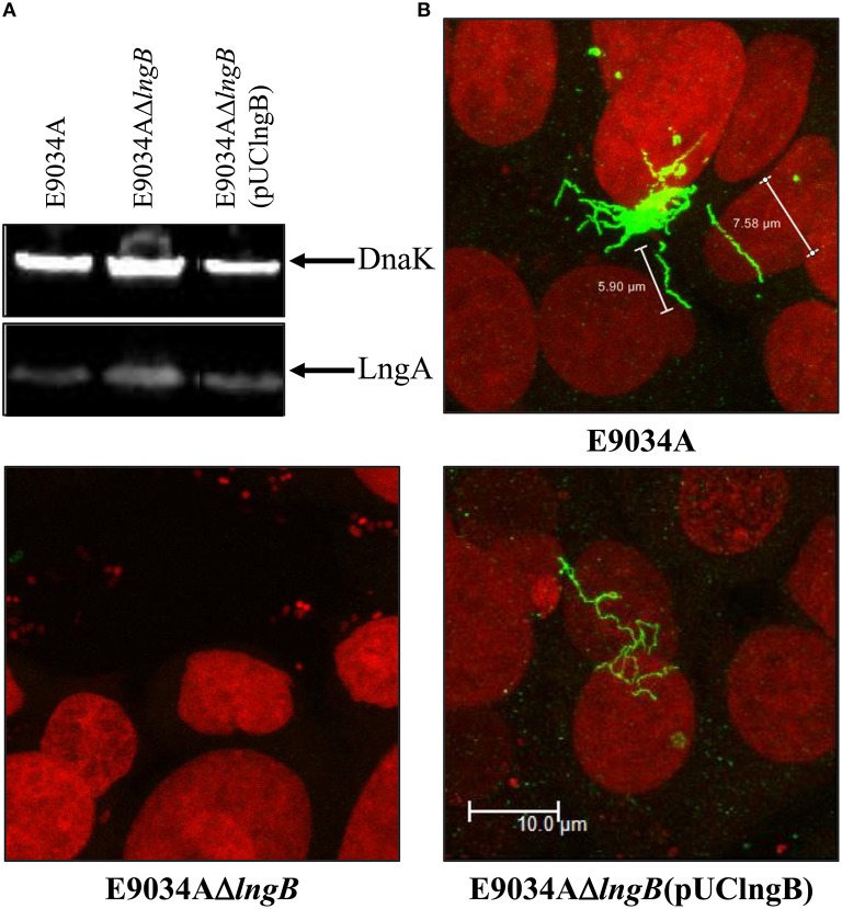
Figure 6.
Complementation of lngB gene in trans restored CS21 expression. (A) Western-blot assay: whole protein extracts were separated by SDS-PAGE, transferred to PVDF membranes, and reacted with anti-CS21 serum and anti-DnaK antibodies. (B) Immunofluorescence assay: eukaryotic and bacterial DNA were stained with propidium iodide (red) and CS21 structures were visualized with anti-CS21 serum and goat anti-rabbit IgG antibodies conjugated with Alexa fluor 488 (green). The confocal microscopy micrographs were taken at a magnification of 63x.

