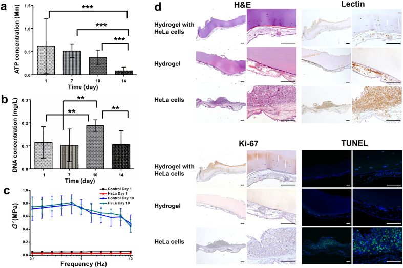
Figure 4. HeLa cell encapsulation and SF hydrogel anti-angiogenic effect.
(a) Cell proliferation and viability measured by (a) ATP assay and (b) DNA quantification, respectively, for SF hydrogel with encapsulated HeLa cells after 1, 7, 10 and 14 days of culture (n = 9). (c) storage modulus for the cancerous cell laden-hydrogels, tested by dynamic mechanical analysis (n = 3). Control Day 1 and Control Day 10: hydrogels without cells encapsulated freshly prepared and 10 days post-preparation; HeLa Day 1 and HeLa Day 10: hydrogels encapsulated with HeLa cells for one day and ten days, respectively. (d) H&E staining, SNA-lectin and Ki-67 immunohistochemical analysis and fluoresence TUNEL assay of the excised section from the CAM study (scale bar: 200 μm). **P < 0.01, ***P < 0.001.

