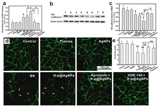
Figure 5. MUA@AgNPs activated plasma increased the paracellular permeability of human retina endothelial cells (HRECs) through regulating adherens junction protein, VE-cadherin.
(a) Monolayer HREC permeability changes upon MUA@ANPs-activated mouse plasma stimulation for 6 h. (b) VE-cadherin expression in HRECs: 1) PBS, 2) 1% (V/V) mouse plasma, 3) 2 μg/mL MUA@AgNPs, 4) 10 μM BK, 5) L-p@AgNPs (0.2 μg/mL MUA@AgNPs activated mouse plasma, 1%, V/V), 6) H-p@AgNPs (2 μg/mL MUA@AgNPs activated mouse plasma, 1%, V/V), 7) 10 μM aprotinin + H-p@AgNPs (2 μg/mL MUA@AgNPs activated mouse plasma, 1%, V/V with 10 μM aprotinin pretreatment), and 8) 2 μM HOE 140 + H-p@AgNPs (HRECs were pretreated with 2 μM HOE 140, then stimulated with 2 μg/mL MUA@AgNPs activated mouse plasma, 1%, V/V). (c) The statistical analysis of the Western blots for VE-cadherin expression; (d) Immunostaining of VE-cadherin on HREC cells treated as indicated. (e) The statistical result of the continuity grade for VE-cadherin expression on HRECs. All results were from three independent experiments, *P < 0.05 and **P < 0.01 by ANOVA.

