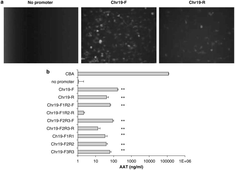
Figure 2.
Chr19 promoter function in human embryonic kidney cells. 293 cells were transfected with plasmid constructs containing different promoter elements from the Chr19 region depicted in Figure 1. (a) The full-length (347 bp) Chr19 element was investigated for promoter activity, paired with gfp, in the forward (F) and reverse (R) orientations. Fluorescent microscopy was performed 24 h after the transfection of 5 μg of plasmid DNA in a 10 cm plate. (b) Promoter activity of the Chr19 region was further dissected using the α-1 antitrypsin (AAT) gene product as the reporter. The primers used for Chr19 region amplification are depicted in Figure 1. The AAT concentration was determined in culture supernatant by ELISA 24 h post-transfection of 5 μg plasmid DNA in a 24-well plate. The chicken β-actin promoter (CBA) was used as a relative control. The results were averaged from four independent experiments and standard deviation is shown. **P<0.01 vs baseline without promoter. ELISA, enzyme-linked immunosorbent assay.

