
Fig. 5.
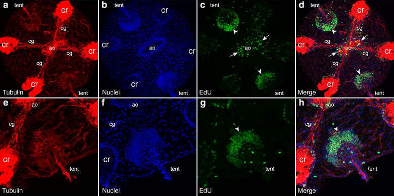
Confocal projections of EdU labeling of Mnemiopsis cydippids (18–24 hpf) to assay for cell proliferation. a–d Confocal projection of aboral portion of cydippid stage labeled with anti-tyrosine tubulin in red (a), which stains the nervous system as well as the cilia of the ciliated groove (cg) and comb rows (cr), Hoechst-stained nuclei in blue (b), EdU-labeled nuclei in green (c) and the merged (d). The apical organ (ao) was at the center, along with the two tentacle bulbs (tent) adjacent. White arrows show EdU labeling in the apical organ floor, and white arrowheads show labeling in the tentacle bulb apparati. e–h Same embryo as in a–d zoomed in close to the tentacle bulb