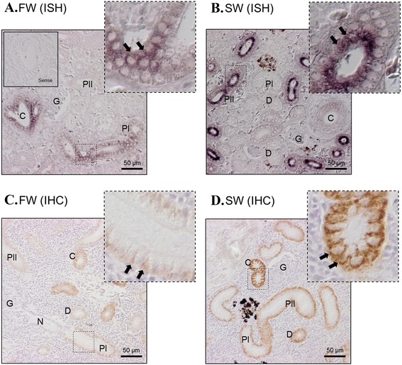
Fig. 8.

In situ hybridization of NKA α1c-1 (a, b) and immunohistochemistry of NKA protein (c, d) in the kidney of eels acclimated to FW and 7 day SW. Positive mRNA signals (arrows) were localized at the proximal tubules (both FW and SW) and collecting tubules (FW > SW). From immunohistochemistry, NKA protein signals were observed at the basolateral side of proximal tubules < distal tubules < collecting tubules. Stronger immunohistochemistry signals was generally found in SW eel compared to FW eel. Top right photos show the structure of the square region in high resolution. Enclosed photos show the negative control of the hybridization incubated with sense probes. G = glomerulus; N = neck; PI = first segment of proximal tubules; PII = second segment of proximal tubules; D = distal tubules; C = collecting tubules
