
Fig. 10.
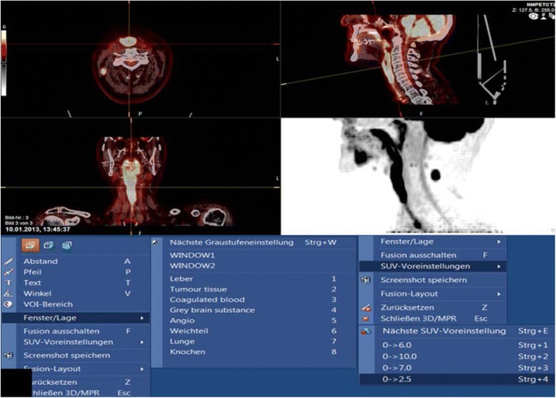
PACS system Sectra Workstation IDS7 Version 17.1.10 with NM Fusion image display allowing demonstration of the volumetric PET-CT data in all three planes and as MIP. The various preset window and SUV settings are shown in the bottom half. The PET images shows the long segment of avid enhancement of the neopharynx in a patient with tumour recurrence (same patient as in Fig. 8) and a lymph node metastasis on the right