
Fig. 6.
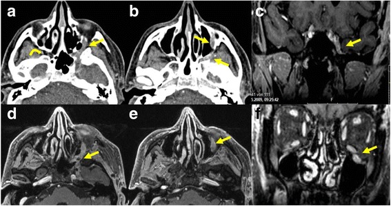
Patient with adenoid cystic carcinoma and perineural extension along the inferior orbital nerve, the pteryopalatine fossa and the maxillary nerve on the left side. Contrast enhanced CT images a, b showing the asymmetric thickened enhancing nerve (arrows) on the left. The curved arrow a shows the normal fat density of the pterygopalatine fossa on the right side. Axial T1-weighted images d, e showing the thickened enhancing nerve (arrows) on the left. Coronal T1-weighted images with fat saturation after injection of gadolinium c showing asymmetrical nerve enhancement (arrow) in the cavernous sinus and f the thickened infraorbital nerve on the left (arrow)