Figure 3.
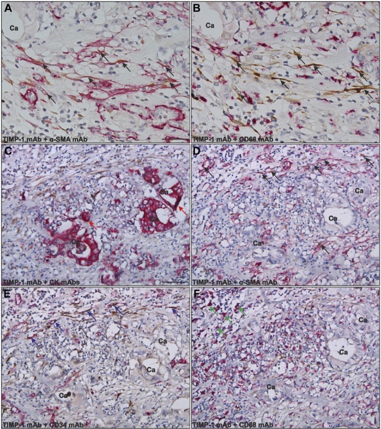
Identification of TIMP-1-positive cells in gastric cancer. Adjacent paraffin-embedded tissue sections were processed for double immunohistochemistry for TIMP-1 and α-SMA (A, D), TIMP-1 and CK (C), TIMP-1 and CD34 (E), or TIMP-1 and CD68 (B, F) using the EnVision G|2 Double System kit from Dako. The TIMP-1 staining was visualized with DAB (brown) and the stainings for α-SMA, CD68, CK, and CD34 were visualized with Permanent Red (magenta). Numerous TIMP-1-positive cells were found at the periphery of the cancer (indicated with Ca) in all samples tested. The vast majority of the TIMP-1-positive cells (black arrows in panels A, B, D) are also positive for α-SMA (black arrows in A, D) but negative for CD68 (B), indicating that these cells are myofibroblasts (panels A and B represent adjacent sections). TIMP-1 positivity is also seen in a few cancer cells (red arrows in panel C), in CD34-positive endothelial cells (blue arrows in panel E), and in a few CD68-positive macrophages (green arrows in panel F). Scale bars: A, B = 30 µm; C–F = 120 µm. Abbreviations: TIMP-1, tissue inhibitor of metalloproteinase-1; α-SMA, α-smooth muscle actin; CK, cytokeratin; DAB, diaminobenzidine; mAb, monoclonal antibody.

