
Fig. 1.
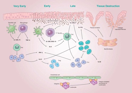
The immunopathogenesis of inflammatory bowel disease (IBD) showing the initial immune response and amplification of the immune response leading to the phenotypic expression of the disease and tissue destruction. Recruitment of leukocytes is mediated by interactions between adhesion molecules known as integrins and the intestinal endothelium. Structural changes to the epithelial barrier result in microbial antigens from the gut lumen entering the host tissue. The initial immune response to the presence of these antigens involves production of interleukin (IL)-7, IL-12, and IL-15, and activation of immune cells, including dendritic cells and macrophages. This results in a T helper (Th) 1 cell response and production of tumor necrosis factor (TNF)-α and interferon (IFN)-γ. The subsequent adaptive immune response results in an amplification of the initial inflammatory response characterized by high levels of IL-6 and IL-23 and the Th17-mediated production of IL-17A, IL-21, and IL-22. The accumulation of myofibroblasts in response to the proliferation of multiple inflammatory cytokines leads to an imbalance in the normal epithelial healing process, resulting in fibrosis and the formation of fistulae. Treg, T regulatory cell.