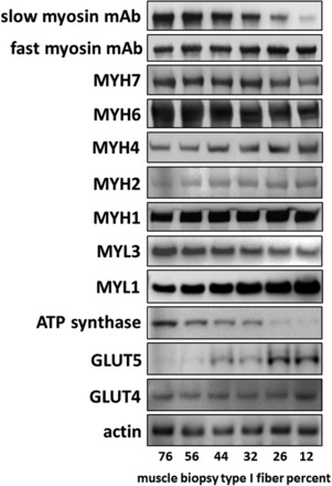
Fig. 2.

Western blots showing protein expression across a range of muscle type I fiber content. Muscle specimen homogenates from 6 subjects with differing muscle fiber content were subjected to PAGE using a 4–12% acrylamide gradient gel with 5 or 20 μg of protein per lane, depending on the antibody to be used. Percent type I fibers was determined using the fast and slow myosin monoclonal antibodies with bright-field images, as described in materials and methods. Specifications of the antibodies are listed in materials and methods. Actin content demonstrated consistent sample protein loading across the spectrum of differing type I fiber content. MYH, myosin heavy chain; MYL, myosin light chain.
