
Fig. 5.
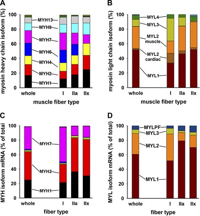
LCM-collected fibers express a mixture of fast and slow myosins. Fiber-specific samples were collected by LCM in triplicate, homogenized, trypsinized, and subjected to MS. A and B: relative amounts of uniquely identified isoform fragments for myosin heavy and light chains. Major isoforms are shown next to the corresponding color. Small amounts of MYH14, MYH15, and MYH16 were detected and are represented as small stacks at the top of the bars in A. Similarly, in B, small amounts of phosphorylatable myosin light chain are displayed as unlabeled blue stacks at the top of the “whole” and IIx bars. In B, MYL2 expression is divided into “MYL2 cardiac” (cross-hatched) and “MYL2 muscle” within the orange color stack. C and D: data from corresponding samples subjected to next-generation sequencing (see materials and methods). Data represent 4 samples obtained by LCM from sections of a muscle specimen from a normal control subject. RNA was isolated from ∼300 transversely cut fibers making up a total cross-sectional area of 2.0 mm2.