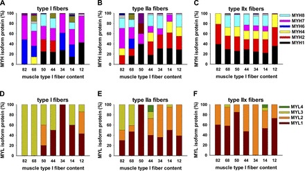
Fig. 6.

LCM-obtained muscle fiber-specific samples from diverse subjects show content of myosin heavy and light chain isoforms in type I, IIa, and IIx fibers. Colored bars show individual subject biopsy fiber type samples analyzed by MS to quantify myosin heavy and light chain isoforms. Data were obtained from slides of muscle biopsies from the same individuals that were used in whole muscle MS studies in Fig. 3.
