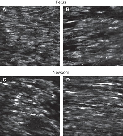
Fig. 1.

Representative micrographs of the endothelium in fetal pulmonary arteries. A–D: average fluo 4 fluorescence signals in endothelial cells of pulmonary arterial segments isolated from fetal sheep exposed to normoxia (A) and prenatal chronic hypoxia (B) and newborn sheep exposed to normoxia (C) and prenatal chronic hypoxia (D). Images represent average intensity from 100 frames for animals exposed to normoxia and 120 frames for animals exposed to hypoxia at 1.28 frames per second. Image brightness was adjusted for display purposes. Images were obtained with a ×40 water-immersion Plan-Apochromat objective. Scale bar = 20 μm.
