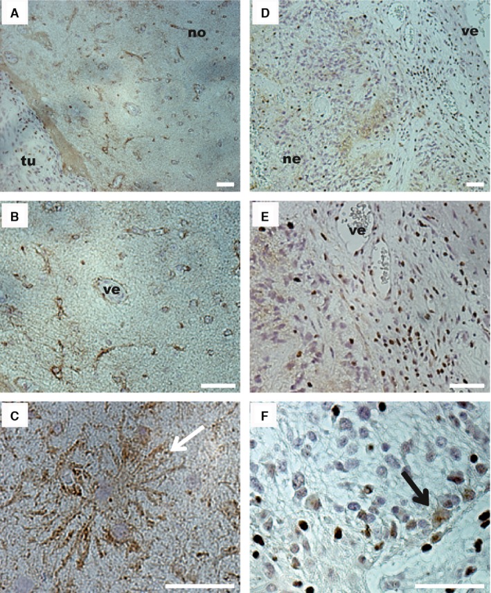
Figure 1.

Cx43 expression observed by immunohistochemistry in pathological and surrounding areas of a grade IV glioma. (A) The surrounding nontumor area (no) exhibited a lower cell density than the tumor area (tu). Surrounding nontumor area (B–C). (B) In this part of the tissue, Cx43 staining (brown) was observed around astrocytes and at the periphery of vessels (ve). (C) At higher magnification, Cx43 staining appeared at the membrane of astrocytes (white arrow) and a more diffuse staining corresponding to the fibrillary background was observed. Tumor area (D–F). (D) Within the tumor, the tissue was disorganized and exhibited high cell density, vascular proliferation (ve) with a thick endothelium, necrotic zones (ne), and a diffuse staining for Cx43 in some parts of the tumor (brown labeling). (E) Surrounding the newly formed blood vessel (ve), Cx43 was not detected. (F) Some cells in the tumor area exhibited Cx43 in the cytoplasm (black arrow). Bar: 50 μm.
