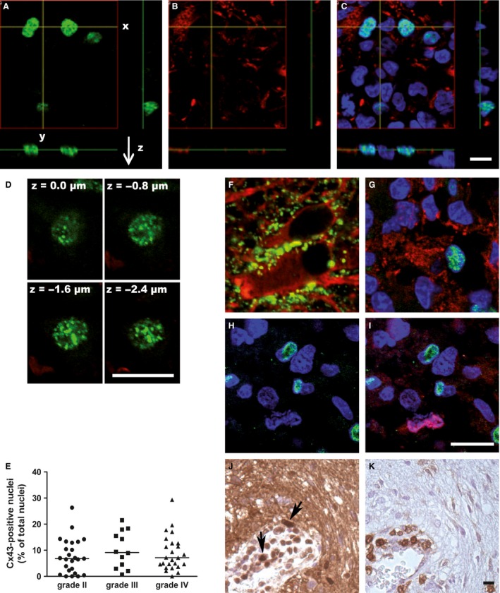
Figure 5.

Presence of a Cx43 nuclear staining in the core of the tumors. (A, B, C) Confocal analysis of the Cx43 staining in 3D (x‐, y‐, and z‐axis). A Cx43 signal (green) was detected in the nucleus (blue) of several cells (A, C). This staining seemed to be detected in cells that did not express a specific astrocyte protein, GFAP (in red; B, C). (D) Confocal analysis of the Cx43 nuclear staining in the nucleus thickness following the z‐axis. The Cx43 signal was shown in successive focal plans within the nucleus. (E) Quantification of the Cx43‐stained nuclei in the different tumor grades (II to IV). No statistically significant difference was shown between the three groups. (F–K) Endeavor of identification of the cell type exhibiting Cx43‐stained nuclei. In the surrounding nontumor area (F), a characteristic Cx43 staining (in green) was shown at the cell–cell contact areas between astrocytes (GFAP, in red). In the core of the tumor (G), a cell exhibiting a Cx43‐stained nucleus (green and blue) appeared GFAP negative, on the contrary of surrounding cells (blue nuclei) that were GFAP positive (red) and did not show Cx43 expression. In the core of the tumor (H, I), costaining was performed for the proliferation nuclear‐marker Ki67 and Cx43. The nucleus of proliferating cells (in red; I) did not colocalize with the Cx43 nuclear staining (in green; H, I). As an accumulation of Cx43 nuclear staining was seen surrounding the newly formed vessels (J; in brown; black arrows), histochemistry assays were made on serial slides for the leukocyte marker CD45 (K; in brown). The areas of high Cx43 nuclear staining appeared correlated with the CD45‐positive cells, especially close to the endothelium. Bar: 50 μm.
