Figure 1.

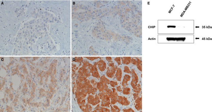
(A‐D) Immunohistochemical findings of CHIP. (A) No staining (score 0), (B) weak staining (score 1), (C) moderate staining (score 2), and (D) strong staining (score 3) for CHIP expression was detected in the cytoplasm of cancer cells. (E) A CHIP protein analysis by western blotting in a breast cancer cell line. The CHIP antibody used in this study clearly detected CHIP proteins in MCF7 cells, but not in MDA‐MB231 cells.
