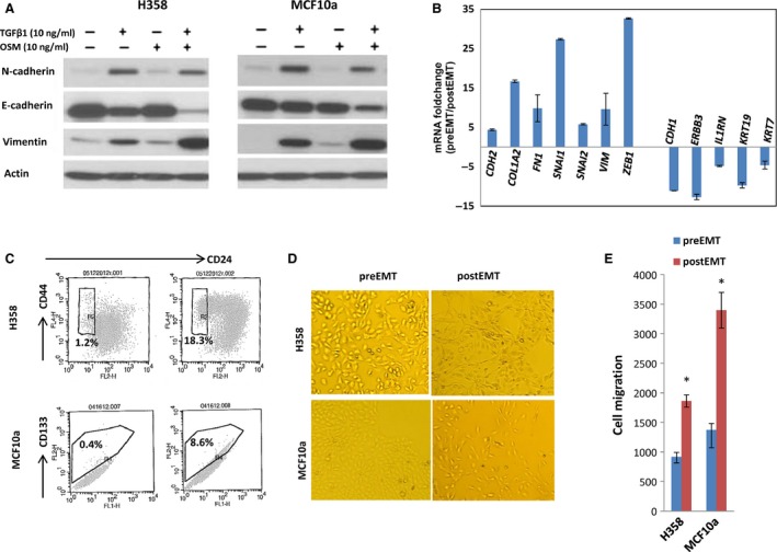
Figure 1.

An optimized TGF‐beta/OSM approach to efficiently induce epithelial mesenchymal transition (EMT) in H358 and MCF10a cells. (A) Western Blot of EMT protein for H358 cells (left) and for MCF10a cells (right). After 7 days of exposure to TGF‐beta/OSM, a clear loss of epithelial biomarker E‐cadherin and increased expression of mesenchymal proteins vimentin and N‐cadherin are shown; (B) RT‐qPCR quantification of 12 important EMT genes in H358 cells treated with TGF‐beta/OSM; (C) TGF‐beta/OSM‐induced CSC‐like cells: CD44+/CD24− subpopulation in H358 cells and CD133+ subpopulation in MCF10a cells. (D) Mesenchymal morphological changes; (E) cell invasiveness. *P < 0.05.
