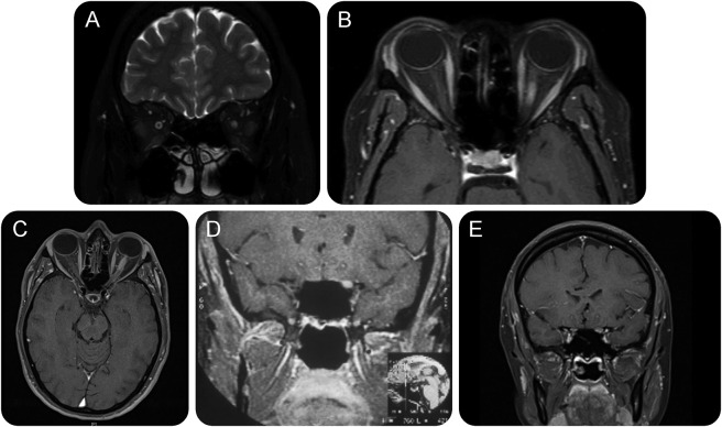
Figure 1. MRI in subacute optic neuritis caused by sarcoidosis.
MRI showing (A) swelling and high signal within the nerve, and (B) enhancement in a patient who presented with a painless subacute optic neuropathy to no perception of light over 5 days. With oral corticosteroid treatment, she recovered to 6/6 (20/20, 1.0). Azathioprine was added and oral steroids withdrawn over 6 months. She has remained well since. (C) T1-weighted axial MRI showing swelling and enhancement of the anterior half of the left optic nerve. (D, E) T1-weighted coronal MRI showing 2 other cases in which the nerve is seen to be swollen and enhancing following administration of contrast. Each case had a subacute optic neuritis with improvement after administration of corticosteroids.

