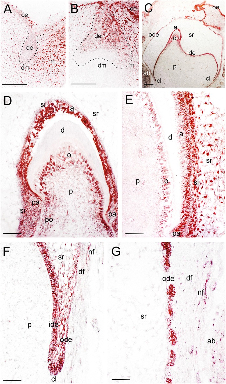
Figure 2.

Distribution of the NGF protein in developing human teeth. (A) NGF immunoreactivity (red color) in a tooth germ at the bud stage. (B) NGF expression in a tooth germ at the early cap stage. (C–G) NGF staining in a tooth germ at the late bell stage. (D–G) Higher magnifications of (C), representing the tip of the cusp area (D), an area of the flanks of the forming tooth crown (E), the cervical loop territory (F), and the dental follicle territory (G). Dotted lines represent the border between the dental epithelial and mesenchymal components. Abbreviations: a, ameloblasts; ab, alveolar bone; cl, cervical loop; d, dentinee; de, dental epithelium; df, dental follicle; dm, dental mesenchyme; ide, inner dental epithelium; m, mesenchyme; nf, nerve fibers; o, odontoblasts; ode, outer dental epithelium; oe, oral epithelium; p, dental pulp; pa, preameloblasts; po, preodontoblasts; si, stratum intermedium; sr, stellate reticulum. Scale bars: (A–C) 100 μm, (D–G) 25 μm.
