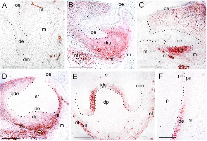
Figure 3.
Expression of p75NTR during tooth development. (A) A tooth germ at the bud stage. Note the p75NTR immunoreactivity (red color) in the nerve fibers (nf) approaching the tooth germ. (B–E) Tooth germs at the cap stage of development. Note that p75NTR staining in mesenchyme is localized in the dental mesenchyme (dm) at the early cap stage (B,C), while the staining is mostly seen in the dental follicle (df) at the late cap stage (D,E). Cells from the inner dental epithelium (ide) start to express p75NTR, while no staining is detected in the dental papilla (dp) at this late stage (E). Note that nerve fibers (nf) surrounding the dental follicle are strongly stained. (F) Higher magnification of the flank area of a tooth germ at the early bell stage of development (Figure 1H). Restricted p75NTR reactivity in undifferentiated cells of the inner dental epithelium. Note that the staining is absent in dental pulp (p). Dotted lines represent the border between the dental epithelial and mesenchymal components. Additional abbreviations: de, dental epithelium; m, mesenchyme; ode, outer dental epithelium; oe, oral epithelium; pa, preameloblasts; po, preodontoblasts; sr, stellate reticulum. Scale bars: 100 μm.

