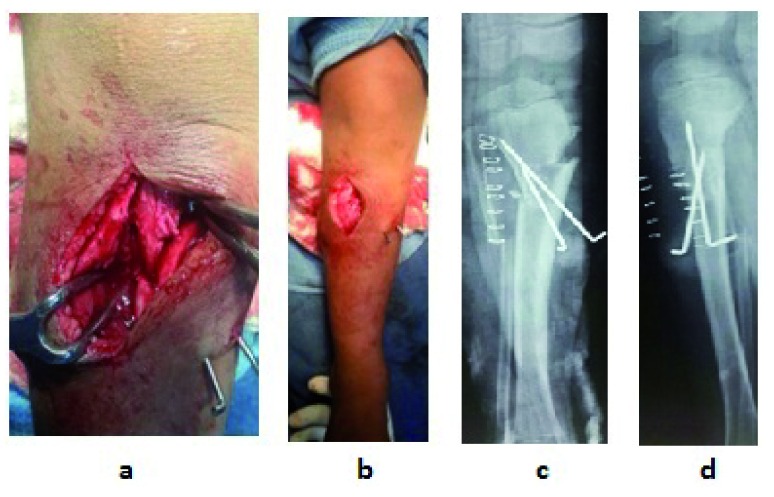
Figure 5. In-operative picture shows the osteotomy site and post-operative X-rays.
a. Close-up view of correction of deformity by Z osteotomy and stabilization with 2 K wires, b. Varus deformity is corrected to normal alignment, c. Immediate post-operative X-ray anterior posterior view, d. Immediate post-operative X-ray lateral view.

