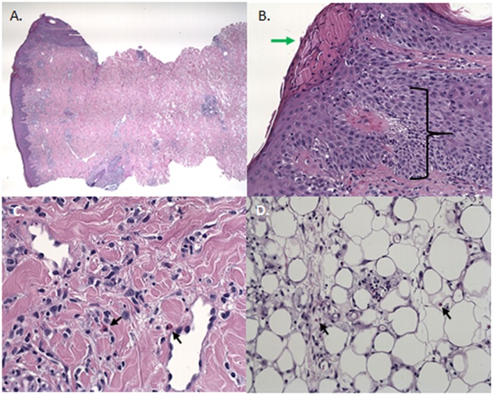
Figure 6.

The histology of a biopsy of an erythematous ISR. A) Overview of biopsy. B) Spongiosis with exocytosis of lymphocytes and parakeratosis with serumcrustae. C) Infiltration with eosinophilic granulocytes. D) Necrosis of the subcutaneous fat tissue and infiltration with eosinophilic granulocytes
