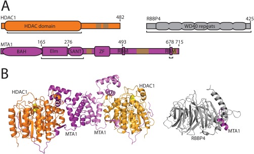
Figure 1.

Schematics of HDAC1, RBBP4 and MTA1. A. Schematics of three components of the NuRD complex. Domains (shown as labelled ovals/boxes) have known structures or the structures of related domains are known. BAH, Elm, SANT, and ZF (zinc finger) are domains of MTA1, whereas RBM refers to RBBP Binding Motifs defined in this article. Brown regions are low complexity sequences. Black underlining refers to protein sections for which crystal structures are available. Colouring is preserved in B. B. Three‐dimensional structures of NuRD subcomplexes. Left: HDAC1‐MTA1ELM‐SANT dimer of dimers (PDB 4BKX). Right: RBBP4‐MTA1675‐686 (PBB 4PBY).
