
Figure 2.
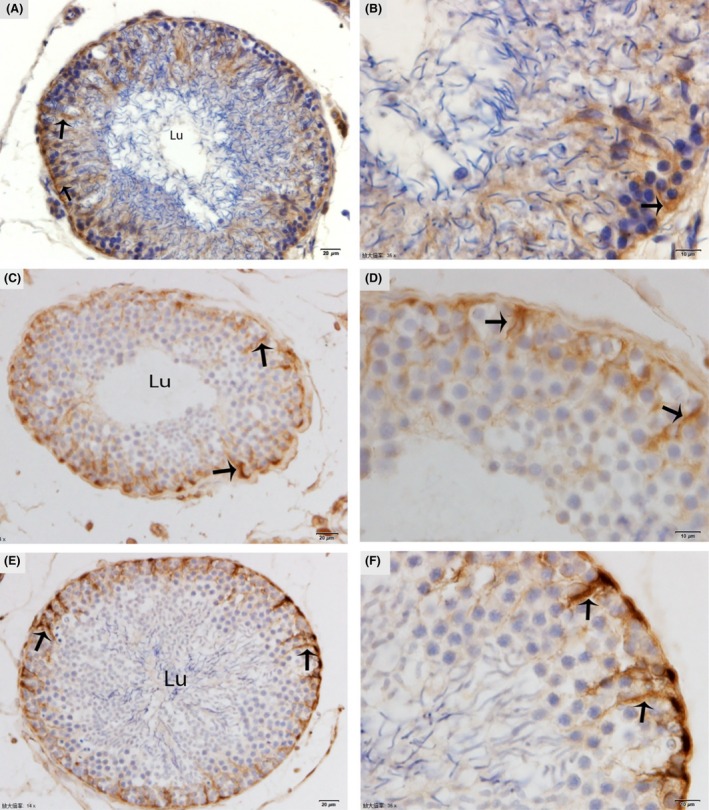
Immunohistochemical localization of vimentin in the testis. The immunoreactivity of Sertoli cells showing weak expression (arrow) in January (A, B), moderate expression (arrow) in May (C, D), and strongly positive expression (arrow) in October (E, F). Lu: lumen. Scale bar = 20 μm (A, C, E) and 10 μm (B, D and F).