
Figure 5.
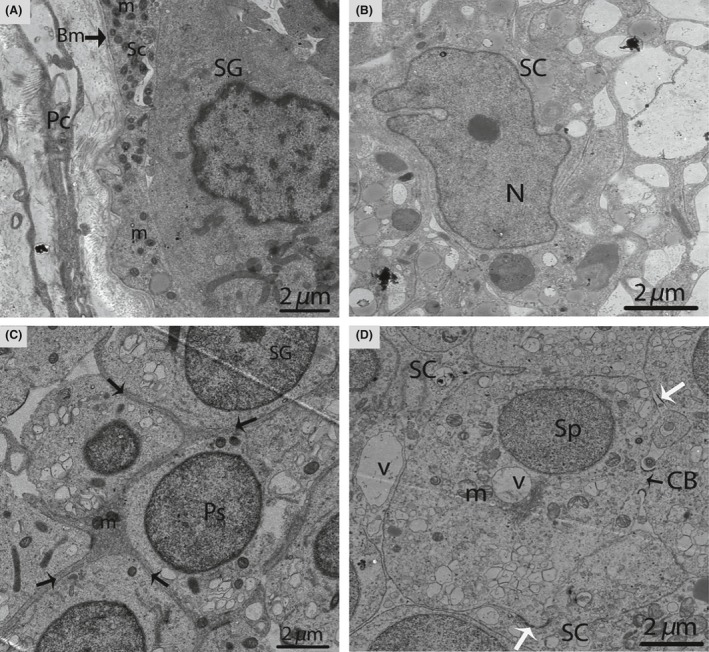
Electron micrograph of seminiferous tubules in October. Sertoli cell exists between the basal membrane and spermatogonia (A). A pear shaped nucleus is observed within the Sertoli cell (B). The thick process of the Sertoli cell (arrow) wraps around the spermatogonia and primary spermatocytes (C). Spermatids are connected by a cytoplasmic bridge and adherens junctions (white arrow) (D). SC, Sertoli cell; Pc, peritubular cell; PS, primary spermatocytes; Sp, spermatid; L, lipid; Bm, basal membrane; SG, spermatogonia; m, mitochondria; CB, cytoplasmic bridge; V, vesicle. Scale bar = 2 μm (A–D).