
Figure 6.
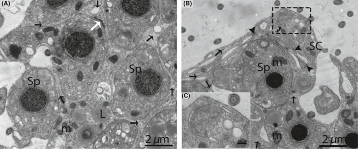
Electron micrograph of the sperm column in October. Sertoli cell (Black arrow) organizes the germ cells into a column of seminiferous epithelia (A). The phagophore (curved arrow) appears within the cytoplasm of a Sertoli cell (B). Illustration indicates the enlarged image of a phagophore (C). Sp, spermatid; SC, Sertoli cell; m, mitochondria; L, lipid; (white arrow) adherens junctions; (arrowhead): tight junctions. Scale bar = 2 μm (A, B) and 200 nm (C).