
Figure 8.
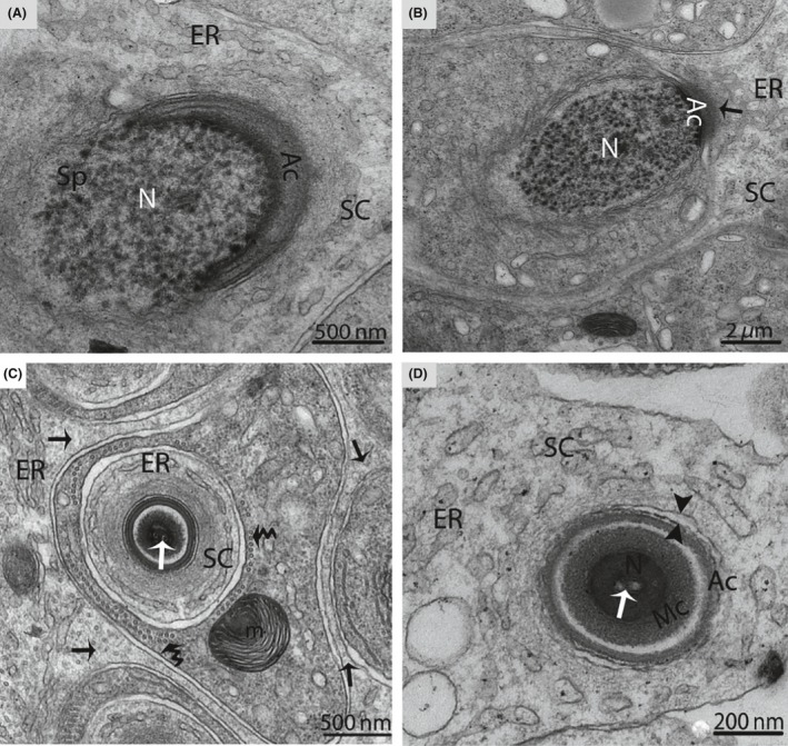
Electron micrograph of a Sertoli cell during spermiogenesis in October. The Sertoli cell (arrow) fully encloses the spermatid during the formation of the acrosomal cap (A). Endoplasmic reticulum secretes a dense material (arrow) near the acrosome (B), and a large number of tubules are observed inside the Sertoli cell (arrow) (C). Opposite arrowheads indicate the cell membrane of the Sertoli cell, which is separated from the spermatid (D). SC, Sertoli cell; Sp, spermatid; Ac, acrosome; N, nucleus; ER, endoplasmic reticulum; m, mitochondria; Mc, manchette; (white arrow): intranuclear canal; (curved arrow): microtubules. Scale bar = 500 nm (A), 2 μm (B), 200 nm (C) and 500 nm (D).