Figure 4.
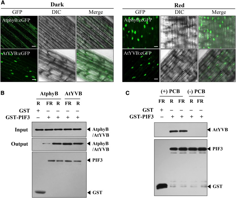
AtYVB is localized in the nucleus and interacts with PIF3 in a light-independent manner. A, Nuclear localization analysis of AtphyB:eGFP and AtYVB:eGFP. Three-day-old dark-grown seedlings were kept in the dark or exposed to R light (5.0 µmol m−2 s−1) for 1 h. DIC, Different interference contrast. Scale bar = 10 μm. B, In vitro protein-protein interaction analysis between AtphyB/AtYVB and PIF3. AtphyB-specific (aN-20) and GST-specific (sc-138) antibodies were used for the detection of AtphyB/AtYVB and PIF3 proteins, respectively. GST was included as a negative control. The phytochrome proteins were expressed in P. pastoris and purified using streptavidin affinity chromatography. PIF3 protein was expressed in E. coli and purified using glutathione affinity chromatography. C, Interaction analysis of PIF3 with apo- and holo-proteins of AtYVB. After preparing ammonium sulfate-precipitated extracts from P. pastoris cells, apo- and holo-proteins of AtYVB were purified without addition of chromophore [(−)PCB] or with the chromophore addition [(+)PCB], respectively.

