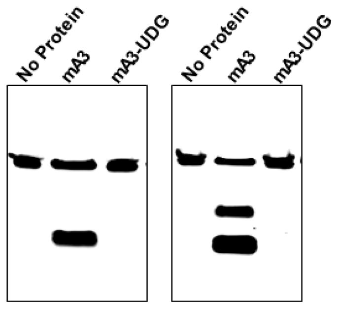
Figure 1. Mouse APOBEC3 was tested with two 48-base oligodeoxynucleotides.

The first (left panel) was composed entirely of A and T residues, except for residues 25– 27, which were TTC, and which was labeled with AlexaFluor 488 at its 5’ end. The oligonucleotide was incubated with no protein (first lane) and with mouse APOBEC3 produced in Sf9 cells (second and third lanes). The second lane shows the results of this incubation, followed by the full assay protocol described here, while the third lane shows the results when UDG was omitted from the assay. The right panel shows a similar assay, but the oligonucleotide contained, in addition to A and T residues, TCCC at residues 21–24 and TTCC at residues 28–31.
