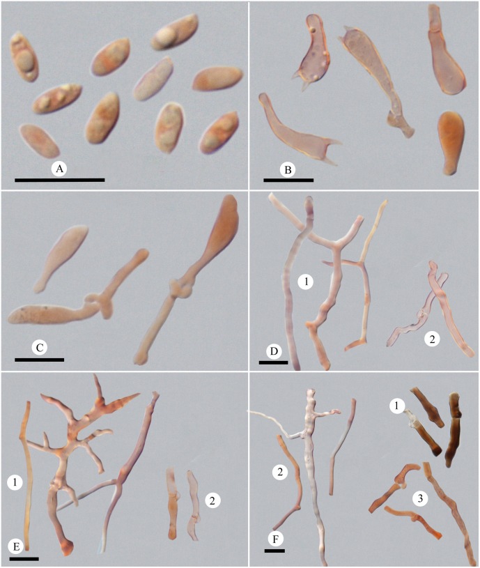
Fig 4. Microscopic structures of Picipes subtropicus.
(A): Basidiospores; (B): Basidia and basidioles; (C): Cystidioles; (D): Hyphae from context, 1 skeleto-binding hyphae, 2 generative hyphae, 3; (E): Hyphae from trama, 1 skeleto-binding hyphae, 2 generative hyphae; (F): Hyphae from stipe, 1 hyphae in cuticle, 2 skeleto-binding hyphae, 3 generative hyphae. Bars = 10 μm.

