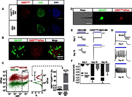
Fig. 1. ChR integration via homologous recombination results in stable expression in ESC and ES-derived MNs and proper light-driven neuronal stimulation.

(A) Membrane-bound expression of tdTomato-tagged ChR2 is observed in transfected ES colonies. Immunostaining for Oct4 expression confirms the pluripotent nature of the transformed cells. Scale bar, 50 μm. DAPI, 4′,6-diamidino-2-phenylindole. (B) Confocal image of a ChR-HBG3–derived neurosphere on day 7 after RA and SAG treatment, showing persistent expression of ChR. (C) FACS data comparing tdTomato::ChR expression of parental (HBG3-MN) and ChR-expressing (ChR-HBG3-MN) cells dissociated from day 7 neurospheres, demonstrating robust expression and minimum silencing after reaching the MN lineage. (D) Dissociated Hb9GFP+/ChRtdTom+ MN plated on a monolayer of cortical glial feeder cells assuming proper neuronal morphology on day 3. The phase contrast image features the patching electrode. Scale bar, 50 μm. (E) Representative trace displaying inward current upon optical stimulation (blue bar) on days 3 and 10 on HBG3-MN and ChRH134R-HBG3-MN. (F) Peak and steady-state inward currents on days 3, 10, and 16 in ChR-HBG3-MN (n = 10). Error bars, SD. (G) Representative current-clamp recordings upon prolonged 1-s optical stimulation displaying AP elicitation on days 3, 10, and 16.
