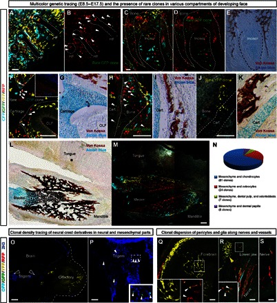
Fig. 7. Cell fate potential of individual neural crest–derived ectomesenchymal clones.

(A to I) Genetic tracing of neural crest–derived progeny in PLP-CreERT2/R26Confetti embryos. Tamoxifen was injected at E8.5, and the analysis of cell types was performed at E17.5. (A and B) Close-up of the region in the anterior face with outlined whisker follicles. Note that cells of the GFP+ rare clone (arrowheads) contribute to both the surrounding mesenchyme and dermal papillae. (C to E) A developing incisor is shown with the dotted line. Note the contribution of the rare GFP+ clone to the peridental mesenchyme, osteoblasts, and the dental mesenchymal compartment. (F and G) Cells of the rare CFP+/RFP+ clone, pointed out by arrowheads, give rise to perichondrial flat cells lining the cartilage, chondrocytes in the olfactory cartilage, and mesenchyme surrounding the olfactory neuroepithelium. (H and I) Arrowheads show cells of the rare GFP+ clone that contribute to cartilage (chondrocytes), trabecular bone (osteocytes), and the surrounding mesenchyme. (J and K) Rare YFP+ clone contributes to the mesenchyme and chondrocytes of the nasal capsule. (L and M) Localized YFP+ and CFP+ clones occupy only a part of a lower jaw and contribute to mandibular bone, Meckel’s cartilage (in the case of the CFP+ clone), and the surrounding loose mesenchyme. (E, G, I, K, and L) Von Kossa staining highlights trabeculae of the facial bones, whereas Alcian blue staining shows cartilage. (N) Diagram representing the occurrence of different cell fate combinations observed within individual clones. (O) Clonal density genetic tracing induced at E8.5 and analyzed at E12.5 in Sox10-CreERT2/R26Confetti embryos. Note the absence of genetically traced cells in the neuroglial compartment despite numerous ectomesenchymal clones present in the location. (P) Example of a GFP+ rare clone (arrowheads) in the neuroglial compartment that does not include ectomesenchymal derivatives. (Q) Part of the network of pericytes in the forebrain is organized by the single yellow clone. NG2 is a pericyte marker, shown in the inset. (R and S) Several traced neural crest–derived glial clones, including a rare YFP+/RFP+ double-colored clone (magnified in the inset), follow the nerve fibers in the lower jaw. For comparison, note the compact ectomesenchymal YFP+ clone in the upper left corner of the image. Scale bars, 200 μm. HF, hair follicle.
