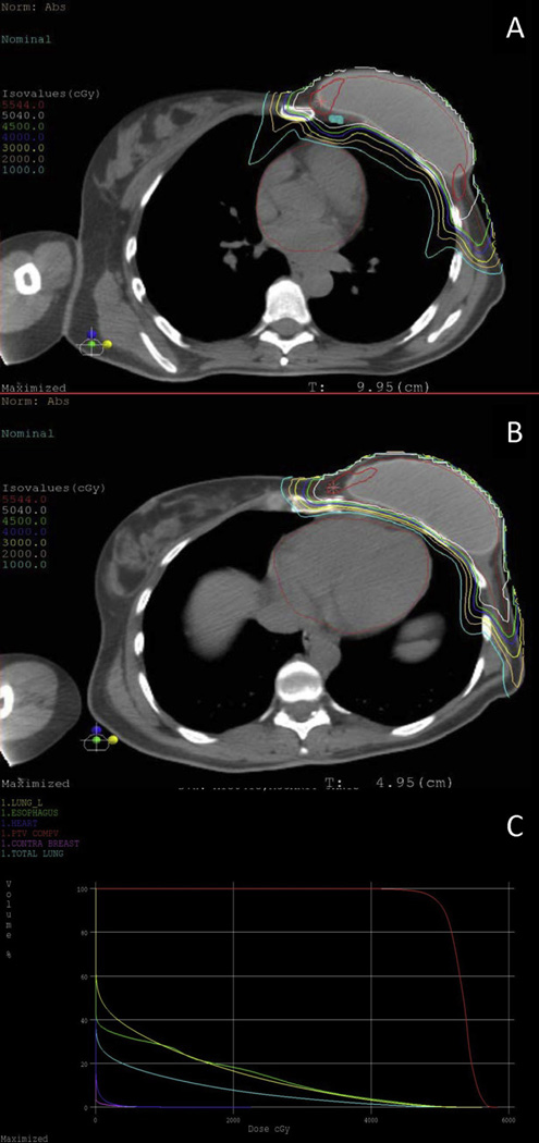
Fig. 4.
Representative dose distribution demonstrating full coverage of (A) internal mammary lymph nodes and (B) heart sparing of a patient treated with postmastectomy proton radiation to the left reconstructed chest wall, axilla, supraclavicular fossa, and internal mammary lymph nodes. (C) Dose–volume histogram.

