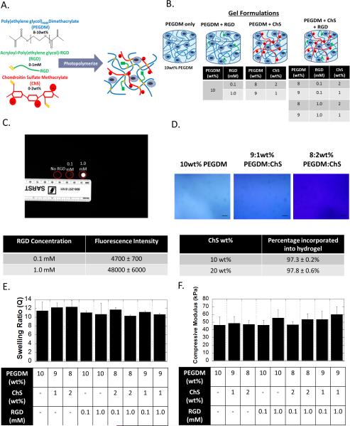
figure 1.
formation and characterization of the biomimetic hydrogels formed from crosslinked poly(ethylene glycol) as the base chemistry and to which (meth)acrylate-functionalized rgd and chondroitin sulfate (chs) are incorporated. a) a schematic showing the monomers and the resulting network structure formed after photopolymerization. b) a schematic of the different hydrogel formulations investigated in this study. c) the incorporation of rgd was confirmed using fluorescently labeled rgd. fluorescent images and the corresponding semi-quantification of fluorescence intensity (n=3) show increased fluorescence with the increasing rgd from 0 to 0.1 to 1 mm. d) the incorporation of chs as confirmed by toluidine blue staining, which stains negatively charged molecules blue. brightfield microscopy images of toluidine blue stained hydrogels with 0, 1, or 2 wt% chs show increased blue staining. scale bar= 100μm. note that peg-only hydrogels exhibit some background staining. the amount of chs incorporated into the hydrogel was also quantitatively assessed (n=3). e) the compressive modulus was measured for the different hydrogel formulations and show no significant differences (n=10, p=0.73). f) the volumetric swelling ratio was measured for the different hydrogel formulations and show no significant differences (n=10, p=0.47). all data are presented as mean with standard deviation parenthetically or as error bars.

