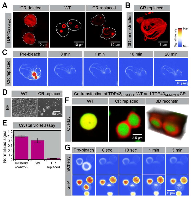
Figure 3. Formation of toxic fibrillar aggregates and multiphase composite assemblies by TDP43 sequence variants.
(A) Images of HEK293T cells transfected with either wild-type (WT) TDP43RRM-mCherry or TDP43RRM-mCherry variants in which the conserved region (CR) was deleted or replaced by a synthetic sequence designed to resemble the flanking IDR (see Figure S1 for details). Cell nuclei are outlined in white. (B) 3D reconstructions of the filigree assemblies formed by the TDP43RRM-mCherry CR replacement variant. (C) No fluorescence recovery is observed when the filamentous assemblies are bleached, suggesting that they represent stable aggregates or polymers. (D) Bright-field (BF) images of TPD43RRM-mCherry droplet- and aggregate-containing cells five days after transfection. (E) Assessment of cell death by crystal violet staining; shown are the crystal violet signals (normalized to the mCherry control) of three independent transfections for each construct (normalized mean ± SD). (F) Co-expression of WT TDP43RRM-GFP (green) and the aggregation-prone TDP43RRM-mCherry CR variant (red) suppresses filament formation; instead a bipartite structure with a mantle around a central core is formed. (G) FRAP analysis of the sub-structured bipartite TDP43 phases.

