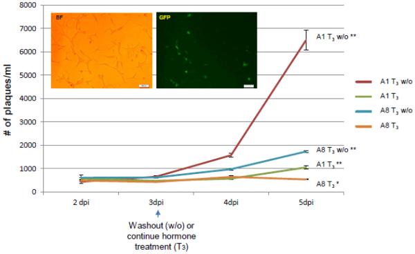
Fig. 5. Assessment of hormone washout effects on viral replication via time course.
Differentiated cells were infected and media supernatants were collected every day for plaque assays. Bright field and fluorescent microscopy, taken at 5 dpi, showed that A8 infected cells maintained healthy morphology. All supernatants were collected at 2, 3, 4, and 5 dpi followed by plaque assays and statistical analyses by ANOVA. The changes in viral release between 2 dpi and 3 dpi are negligible and no significant differences were observed among all treatments. At 4 dpi the viral release of washout samples started to rise significantly in comparison to hormone treated ones, which showed no significant increase. At 5 dpi viral release from all samples showed significant increase (symbolized as **) except T3 treated A8 infected culture (orange line), which still maintained no significanT increase (denoted as *). The RNA samples from infected cells at 5 dpi were purified for subsequent experiments described in Fig. 6.

