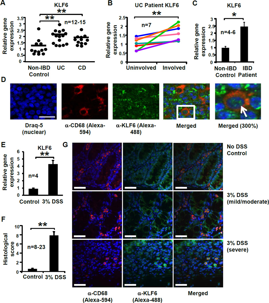
Figure 1. KLF6 is upregulated in inflamed intestinal tissues and macrophages from human and experimental IBD subjects.
(A) Colon tissues from non-IBD control subjects or UC or CD patients were homogenized, cDNA prepared, and analysis of KLF6 gene expression performed by qPCR; **p≤0.001, n=12–15/group. (B) Paired biopsies from uninvolved and involved regions of UC patient colon tissue were analyzed for KLF6 gene expression by qPCR. Fold changes in involved samples were calculated relative to uninvolved (baseline) samples and normalized to expression of SHDA. Lines represent individual patients; **p≤0.03, n=7). (BC) Monocyte-derived macrophages from non-IBD control subjects or IBD patients were analyzed for KLF6 gene expression by qPCR; **p≤0.01, n=4–6/group. (D) KLF6-expressing myeloid cells were detected in human mesenteric lymph nodes by immunofluorescent confocal microscopy. Formalin-fixed, paraffin-embedded mesenteric lymph node tissues were stained with antibodies specific to CD68 (myeloid cell marker, imaged with Alexa 594 as red) and KLF6 (imaged with Alexa 488 as green). Nuclei are stained with Draq5 (imaged as blue); n=3 independent experiments. Scale bar = 100µm; white arrow indicates representative KLF6-expressing macrophage. (E) Macrophages from 3% DSS-fed mice express elevated levels of Klf6. Peripheral blood-derived macrophages from WT mice were analyzed for Klf6 gene expression by qPCR; Graphs show mean ± SEM; **p≤0.001, n=4/group. (F) Histological colon inflammation was assessed by a blinded pathologist following 5 days of 3% DSS. Score consists of active, chronic, and transmural inflammation; **p≤0.001, n=8–23 mice/group. (G) KLF6-expressing myeloid cells are observed in colon tissue from DSS-fed mice by confocal microscopy. Formalin-fixed, paraffin-embedded colon tissues from indicated mice were stained by immunofluorescent confocal microscopy using antibodies specific to CD68 and KLF6, as described above (C). Nuclei were stained with Draq5. Scale bar = 34µm. Images are representative of 3 separate experiments.

