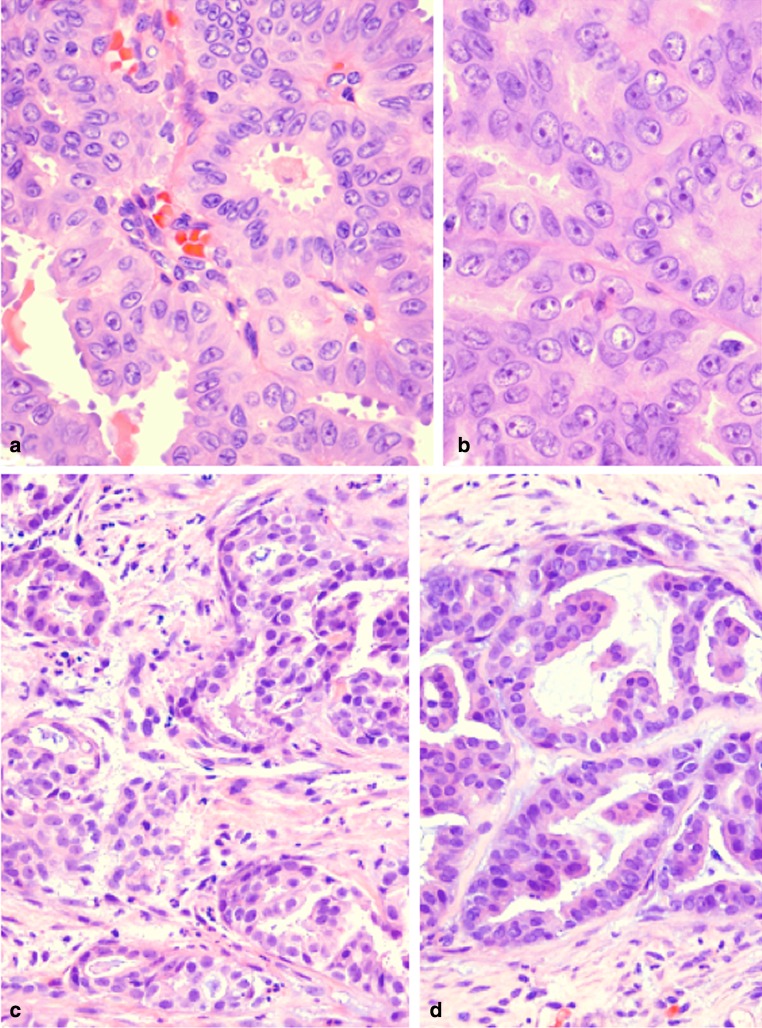
Fig. 3.
a MASC of thyroid (2007). Apocrine-like capitation by lining cells is present in this field (H&E stain). (H&E stain). b MASC of thyroid (2007). Prominent single, centrally placed nucleoli and numerous nuclear grooves are a common feature in all architectural patterns of the tumor (H&E stain). c Metastatic MASC to deltoid muscle (2014). The tumor comprises nests of eosinophilic cells embedded in fibrotic stroma. There is no evidence of transformation to a higher grade (H&E stain). d Metastatic MASC to soft tissue of lower leg (2015). The tumor continues to show similar histology to the original tumor without dedifferentiation (H&E stain)

