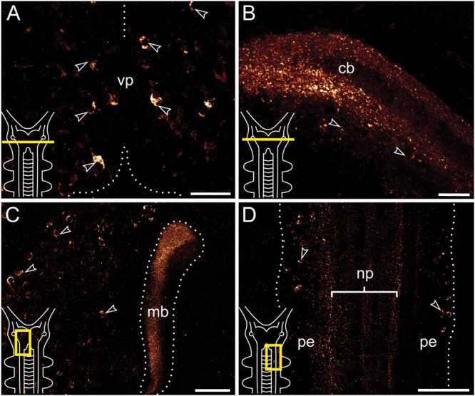
FIGURE 3.

Immunolocalization of arthropsin in the nervous system of the onychophoran Euperipatoides rowelli Vibratome sections. Confocal micrographs. Insets in the lower left corners illustrate sectional planes (yellow bars). (A) Cross section of the ventral portion of the brain. Dorsal is up. Note the ventral group of somata in the protocerebrum (arrowheads). (B) Cross section of the central body. Dorsal is up. Note the outer and inner fiber layers and the associated immunoreactive somata (arrowheads). (C) Horizontal section of the mushroom body. Anterior is up, median is left. Note prominent immunoreactivity in the inner lobe of the mushroom body and numerous immunoreactive somata in the medioventral region of the protocerebrum (arrowheads). (D) Horizontal section of the ventral nerve cord. Anterior is up, median is left. Note immunoreactive fiber bundles within the nerve cord neuropil with associated somata in the perikaryal layer (arrowheads). Abbreviations: cb, central body; mb, mushroom body; np, nerve chord neuropil; pe, perikaryal layer; vp, ventral protocerebrum. Scale bars: 25 μm (A,B), 50 μm (C), 100 μm (D).
