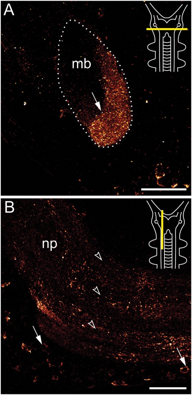
FIGURE 4.

Immunolocalization of arthropsin in the nervous system of the onychophoran Euperipatoides rowelli. Vibratome sections of heads. Confocal micrographs. Dorsal is up in all images. Insets in the upper right corners illustrate the position of sectional planes (yellow bars). (A) Cross section of the mushroom body. Note the presence of immunoreactive fibers in the inner/median lobe (arrow). (B) Sagittal section of anterior nerve cord. Note the immunoreactive somata (arrows) within the perikaryal region and fibers within the nerve cord neuropil (arrowheads). Abbreviations: mb, mushroom body; np, nerve cord neuropil. Scale bars: 50 μm (A,B).
