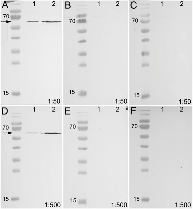
FIGURE 7.

Western blot of unblocked and blocked arthropsin antisera using recombinant expressed protein extracted from Escherichia coli. Truncated Er-arthropsin (20 kDa) fused with MBP (42,5 kDa) was used for all experiments. Lane 1 was loaded with the bacterial cell lysate of Escherichia coli and lane 2 with the purified Er-arthropsin-MBP fusion protein in all images. (A) Unblocked anti-arthropsin antibody (dilution 1:50). Note the two distinct bands at 63 kDa (arrow). (B) Blocked antibody with twice (2x) the amount of the synthetic peptide (1:50). (C) Blocked antibody with 10-fold (10x) the amount of the synthetic peptide. (D) Unblocked anti-arthropsin antibody (1:500). Note the two distinct bands at 63 kDa (arrow). (E) Blocked antibody with twice (2x) the amount of the synthetic peptide (1:500). (F) Blocked antibody with 10-fold (10x) the amount of the synthetic peptide (1:500). Note the absence of specific bands in all experiments with blocked anti-arthropsin antibody (B,C,E,F).
