
FIGURE 5.
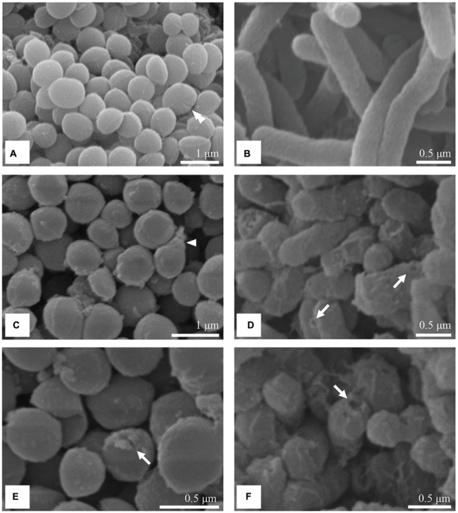
SEM micrographs of untreated and treated S. aureus and S. Enteritidis cells. Untreated cells of S. aureus (A) and S. Enteritidis (B) were intact with regular shape and surface. After treatment with the MIC of the methanol extract of C. versicolor, S. aureus cells looked misshapen (C) and some had holes in their cell wall (E). Treated S. Enteritidis cells appeared shorter, aggregated with dimples and blisters on their surface (D) and ruptured cell envelope (F).